

Wong, Ivy Tsz-Lo; Bailey, Chris; Wu, Sihan; Henssen, Anton G; Cravatt, Benjamin F; Chen, Zhijian J; Bafna, Vineet; Jamal-Hanjani, Mariam; Swanton, Charlie; Chang, Howard Y; Mischel, Paul S
A recipe for chaos: Extrachromosomal DNA and the hallmarks of cancer Journal Article
In: Cell, vol. 189, no. 8, pp. 2307–2321, 2026, ISSN: 1097-4172.
@article{pmid41997126,

title = {A recipe for chaos: Extrachromosomal DNA and the hallmarks of cancer},

author = {Ivy Tsz-Lo Wong and Chris Bailey and Sihan Wu and Anton G Henssen and Benjamin F Cravatt and Zhijian J Chen and Vineet Bafna and Mariam Jamal-Hanjani and Charlie Swanton and Howard Y Chang and Paul S Mischel},

doi = {10.1016/j.cell.2026.03.011},

issn = {1097-4172},

year  = {2026},

date = {2026-04-01},

journal = {Cell},

volume = {189},

number = {8},

pages = {2307--2321},

abstract = {Some aggressive cancers exhibit a level of rapid genome change and therapy resistance that is difficult to explain. Research over the past decade has shown that extrachromosomal DNA (ecDNA) can be the cause. When oncogenic genetic elements untether from chromosomes and no longer follow Mendelian inheritance, genomic chaos and accelerated evolution ensue, generating unique ecDNA biology and non-traditional therapeutic vulnerabilities distinct from traditional mutation-targeting approaches. Here, we put forward a holistic view where ecDNA is integrated into the broader Hallmarks of Cancer framework to better understand the problem and chart a path forward.},

keywords = {},

pubstate = {published},

tppubtype = {article}

}
Werr, Lisa; Boland, Jana; Petersen, Josephine; Iglesias, Fiorella; Höppner, Stefanie; Bartenhagen, Christoph; Rosswog, Carolina; Hellmann, Anna-Maria; Kahlert, Yvonne; Hemstedt, Nadine; Ibruli, Nadliv; Dammert, Marcel A; Decarolis, Boris; Werner, Jan-Michael; Malchers, Florian; Schramm, Kathrin; Witt, Olaf; Beiske, Klaus H; Rognlien, Anne Gro Wesenberg; Gunnes, Maria Winther; Langenberg, Karin P; Molenaar, Jan; Bernkopf, Marie; Taschner-Mandl, Sabine; Hughes, Debbie; George, Sally L; Chesler, Louis; Schulte, Johannes H; Barone, Giuseppe; Capasso, Mario; Surrey, Lea F; Bagatell, Rochelle; Masliah-Planchon, Julien; Schleiermacher, Gudrun; Grüll, Holger; Westermann, Frank; Schultheis, Anne M; Büttner, Reinhard; Henssen, Anton G; Eggert, Angelika; Peifer, Martin; Shukla, Neerav N; Simon, Thorsten; Hero, Barbara; Reinhardt, H Christian; Thomas, Roman K; Fischer, Matthias
Mutated FGFR1 is an oncogenic driver and therapeutic target in high-risk neuroblastoma Journal Article
In: J Clin Invest, vol. 136, no. 7, 2026, ISSN: 1558-8238.
@article{pmid41678281,

title = {Mutated FGFR1 is an oncogenic driver and therapeutic target in high-risk neuroblastoma},

author = {Lisa Werr and Jana Boland and Josephine Petersen and Fiorella Iglesias and Stefanie Höppner and Christoph Bartenhagen and Carolina Rosswog and Anna-Maria Hellmann and Yvonne Kahlert and Nadine Hemstedt and Nadliv Ibruli and Marcel A Dammert and Boris Decarolis and Jan-Michael Werner and Florian Malchers and Kathrin Schramm and Olaf Witt and Klaus H Beiske and Anne Gro Wesenberg Rognlien and Maria Winther Gunnes and Karin P Langenberg and Jan Molenaar and Marie Bernkopf and Sabine Taschner-Mandl and Debbie Hughes and Sally L George and Louis Chesler and Johannes H Schulte and Giuseppe Barone and Mario Capasso and Lea F Surrey and Rochelle Bagatell and Julien Masliah-Planchon and Gudrun Schleiermacher and Holger Grüll and Frank Westermann and Anne M Schultheis and Reinhard Büttner and Anton G Henssen and Angelika Eggert and Martin Peifer and Neerav N Shukla and Thorsten Simon and Barbara Hero and H Christian Reinhardt and Roman K Thomas and Matthias Fischer},

doi = {10.1172/JCI189152},

issn = {1558-8238},

year  = {2026},

date = {2026-04-01},

journal = {J Clin Invest},

volume = {136},

number = {7},

abstract = {Fibroblast growth factor receptor 1 (FGFR1) is recurrently mutated at p.N546 in neuroblastoma. We examined whether mutant FGFR1 is an oncogenic driver, a predictive biomarker, and an actionable vulnerability in this malignancy. FGFR1 mutations at p.N546 were associated with high-risk disease and rapid tumor progression, resulting in dismal outcome for these patients. Ectopic expression of FGFR1N546K induced constitutive downstream signaling and IL-3-independent growth in Ba/F3 cells, indicating oncogene-addicted proliferation. In FGFR1N546K;MYCN transgenic mice, neuroblastoma developed within the first days of life, with fatal outcome within 3 weeks, reflecting the devastating clinical phenotypes of patients with FGFR1-mutant, high-risk neuroblastoma. Treatment with FGFR inhibitors impaired proliferation and pathway activation in FGFR1N546K-expressing Ba/F3 and patient-derived FGFR1N546K-mutant neuroblastoma cells and inhibited tumor growth in FGFR1N546K;MYCN transgenic mice and in a chemotherapy-resistant, patient-derived xenograft mouse model. In addition, partial regression of FGFR1N546K-mutant tumor lesions occurred upon treatment with the FGFR inhibitor futibatinib and low-intensity chemotherapy in a patient with refractory neuroblastoma. Together, our data demonstrate that FGFR1N546K is a strong oncogenic driver in neuroblastoma associated with failure of current standard chemotherapy and suggest potential clinical benefit of FGFR-directed therapies in patients with high-risk mutant FGFR1.},

keywords = {},

pubstate = {published},

tppubtype = {article}

}
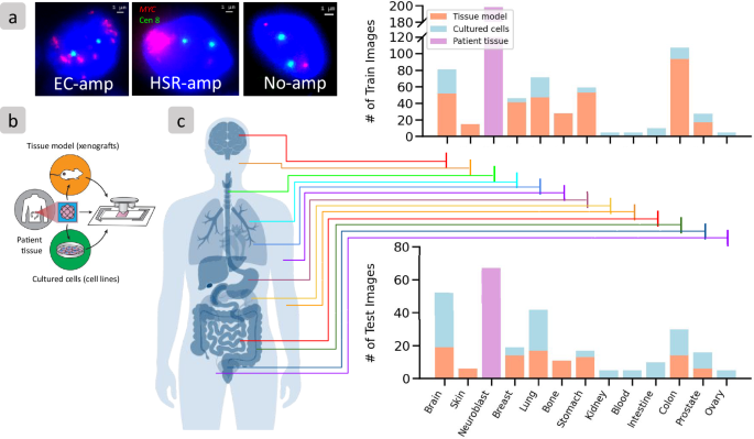
Prasad, Gino; Rajkumar, Utkrisht; Curtis, Ellis J; Wong, Ivy Tsz-Lo; Yan, Xiaowei; Zhang, Shu; Brückner, Lotte; Turner, Kristen; Wiese, Julie; Wahl, Justin; Hemmati, Homa; Wu, Sihan; Theissen, Jessica; Fischer, Matthias; Chang, Howard Y; Henssen, Anton G; Mischel, Paul S; Bafna, Vineet
Accurate prediction of ecDNA in interphase cancer cells using deep neural networks Journal Article
In: Commun Biol, 2026, ISSN: 2399-3642.
@article{pmid41965931,

title = {Accurate prediction of ecDNA in interphase cancer cells using deep neural networks},

author = {Gino Prasad and Utkrisht Rajkumar and Ellis J Curtis and Ivy Tsz-Lo Wong and Xiaowei Yan and Shu Zhang and Lotte Brückner and Kristen Turner and Julie Wiese and Justin Wahl and Homa Hemmati and Sihan Wu and Jessica Theissen and Matthias Fischer and Howard Y Chang and Anton G Henssen and Paul S Mischel and Vineet Bafna},

doi = {10.1038/s42003-026-09982-4},

issn = {2399-3642},

year  = {2026},

date = {2026-04-01},

journal = {Commun Biol},

abstract = {Oncogene amplification is a key driver of cancer pathogenesis and is often mediated by extrachromosomal DNA (ecDNA). EcDNA amplifications are associated with increased pathogenicity of cancer and poorer outcomes for patients. EcDNA can be detected accurately using fluorescence in situ hybridization (FISH) when cells are arrested in metaphase. However, the majority of cancer cells are non-mitotic and must be analyzed in interphase, where it is difficult to discern extrachromosomal amplifications from chromosomal amplifications. Thus, there is a need for methods that accurately predict oncogene amplification status from interphase cells.We present interSeg, a deep learning-based tool to cytogenetically classify oncogene amplification status as extrachromosomally amplified (EC-amp), intrachromosomally amplified (HSR-amp), or not amplified, from interphase FISH images. We trained and validated interSeg on 652 images (40,446 nuclei). Tests on 215 cultured cell and tissue model images (9,733 nuclei) showed 89% and 97% accuracy at the nuclear and sample levels, respectively. The neuroblastoma patient tissue hold-out set (67 samples and 1,937 nuclei) also revealed 97% accuracy at the sample level in detecting the presence of focal amplification. In experimentally and computationally mixed images, interSeg accurately predicted the level of heterogeneity. The results showcase interSeg as an important method for analyzing oncogene amplifications.},

keywords = {},

pubstate = {published},

tppubtype = {article}

}
Margotte, Luna; Henssen, Anton; Thorwarth, Anne; Eggert, Angelika; Scheer, Monika
Number of affected lymph nodes predicts outcome in extremity rhabdomyosarcoma Journal Article
In: Eur J Surg Oncol, vol. 52, no. 3, pp. 110535, 2026, ISSN: 1532-2157.
@article{pmid41666515,

title = {Number of affected lymph nodes predicts outcome in extremity rhabdomyosarcoma},

author = {Luna Margotte and Anton Henssen and Anne Thorwarth and Angelika Eggert and Monika Scheer},

doi = {10.1016/j.ejso.2025.110535},

issn = {1532-2157},

year  = {2026},

date = {2026-03-01},

journal = {Eur J Surg Oncol},

volume = {52},

number = {3},

pages = {110535},

abstract = {BACKGROUND: Rhabdomyosarcoma (RMS) of the extremity has poor outcomes due to its high potential for lymphatic and haematogenic spread.nnMETHODS: Data were selected from the SEER17 registry 2000-2020 for non-pleomorphic extremity rhabdomyosarcoma (all ages and stages).nnRESULTS: 473 cases were identified, 181 at upper and 292 at lower extremity. Median age was 14years. Histology was alveolar in 250. The median tumor size was 7 cm. 144 tumors were localized, 131 regional and 178 distant (20 unknown). Median follow-up for 229 survivors was 8.17years. 5-year-DSS and OS was 49.7 % ± 4.9(95 % CI) and 46.3 % ± 4.9, respectively. A total of 169 patients underwent pathological examination of regional lymph nodes. In 51, one node was examined, in 24 two and in 63 ≥ 3 nodes were examined. Overall, 85 patients had pathologically positive nodes: 36 had one positive regional lymph node, 12 had two and 15 had ≥3. When adjusted for age, histology, size and stage, involvement of ≥3 lymph nodes was independently associated with worse survival. This observation was also applicable to the Cox regression analysis, which exclusively considered the 275 cases of localized/regional stage.nnCONCLUSION: The number of regional lymph nodes affected correlates with survival, suggesting the extent of lymph node involvement as a new and crucial predictive factor. Further analysis of the extent of regional lymph node involvement is advisable. This includes identifying the most effective methods for detecting affected nodes and establishing how many nodes need to be examined to obtain representative results.},

keywords = {},

pubstate = {published},

tppubtype = {article}

}
Hebron, Katie E; Odeniyide, Patience; Wei, Yun; Gryder, Berkley E; Barr, Frederic G; Casey, Dana L; Chen, Eleanor Y; Crompton, Brian D; Cruz, Filemon S Dela; Durbin, Adam D; Ford, Heide L; Gatz, Susanne A; Hatley, Mark E; Henssen, Anton G; Hettmer, Simone; Houghton, Peter J; Kendall, Genevieve C; Khan, Javed; Lupo, Philip J; Patel, Anand G; Pomella, Silvia; Rota, Rossella; Schito, Marco; Schoot, Reineke A; Shern, Jack F; Stanton, Benjamin Z; Stewart, Elizabeth A; Swindlehurst, Cathy A; Thomas, Craig J; Vakoc, Christopher R; Vaseva, Angelina V; Venkatramani, Rajkumar; Wexler, Leonard H; Yustein, Jason T; Hammond, Sharon; Heske, Christine M; Langenau, David M; Linardic, Corinne M; Ignatius, Myron S; Yohe, Marielle E
Biological Advances and Current Challenges for Pediatric Rhabdomyosarcoma Journal Article
In: Cancers (Basel), vol. 18, no. 6, 2026, ISSN: 2072-6694.
@article{pmid41899494,

title = {Biological Advances and Current Challenges for Pediatric Rhabdomyosarcoma},

author = {Katie E Hebron and Patience Odeniyide and Yun Wei and Berkley E Gryder and Frederic G Barr and Dana L Casey and Eleanor Y Chen and Brian D Crompton and Filemon S Dela Cruz and Adam D Durbin and Heide L Ford and Susanne A Gatz and Mark E Hatley and Anton G Henssen and Simone Hettmer and Peter J Houghton and Genevieve C Kendall and Javed Khan and Philip J Lupo and Anand G Patel and Silvia Pomella and Rossella Rota and Marco Schito and Reineke A Schoot and Jack F Shern and Benjamin Z Stanton and Elizabeth A Stewart and Cathy A Swindlehurst and Craig J Thomas and Christopher R Vakoc and Angelina V Vaseva and Rajkumar Venkatramani and Leonard H Wexler and Jason T Yustein and Sharon Hammond and Christine M Heske and David M Langenau and Corinne M Linardic and Myron S Ignatius and Marielle E Yohe},

doi = {10.3390/cancers18060888},

issn = {2072-6694},

year  = {2026},

date = {2026-03-01},

journal = {Cancers (Basel)},

volume = {18},

number = {6},

abstract = {Despite comprehensive and multi-modal therapy, outcomes for children and adolescents with rhabdomyosarcoma (RMS) have plateaued over the past four decades. This is not for a lack of progress in the basic and translational studies of RMS. Indeed, advances in animal models and/or patient tissue sample acquisition and analysis have improved our understanding of RMS biology. Large-scale sequencing efforts have generated transcriptomic, genomic, and epigenomic datasets that highlight the heterogeneity of RMS and have the potential to improve prognostication and the application of precision medicine in patients with RMS. However, few of these discoveries have been clinically translated, and limitations to the accessibility, uniformity, and application of these new models and datasets hinder their utility. Here, we discuss how advances in understanding RMS biology, optimization of preclinical models, and strategies for translating basic science discoveries to the clinic can potentially improve outcomes for patients with RMS.},

keywords = {},

pubstate = {published},

tppubtype = {article}

}
Gatz, Susanne A; Glade-Bender, Julia; Pearson, Andrew D J; Ortiz, Michael V; Bernardi, Ronald; Chesler, Lou; Clifford, Steve; Cohen-Gogo, Sarah; Cuesta, Esther De La; de Rojas, Teresa; Durinck, Kaat; Federico, Sara; Fox, Elizabeth; George, Sally; Gounaris, Ioannis; Henssen, Anton George; Irwin, Meredith; Kool, Marcel; Lau, Alan; Nysom, Karsten; Pappo, Alberto; Pennock, Gregory K; Pfister, Stefan M; Scobie, Nicole; Slotkin, Emily K; Smith, Malcolm; Speleman, Frank; Stewart, Elizabeth A; Weigel, Brenda J; Vassal, Gilles
Pathway for the Development of ATR Inhibitors in Pediatric Malignancies: An ACCELERATE Multistakeholder Analysis Journal Article
In: JCO Precis Oncol, vol. 10, pp. e2500642, 2026, ISSN: 2473-4284.
@article{pmid41538758,

title = {Pathway for the Development of ATR Inhibitors in Pediatric Malignancies: An ACCELERATE Multistakeholder Analysis},

author = {Susanne A Gatz and Julia Glade-Bender and Andrew D J Pearson and Michael V Ortiz and Ronald Bernardi and Lou Chesler and Steve Clifford and Sarah Cohen-Gogo and Esther De La Cuesta and Teresa de Rojas and Kaat Durinck and Sara Federico and Elizabeth Fox and Sally George and Ioannis Gounaris and Anton George Henssen and Meredith Irwin and Marcel Kool and Alan Lau and Karsten Nysom and Alberto Pappo and Gregory K Pennock and Stefan M Pfister and Nicole Scobie and Emily K Slotkin and Malcolm Smith and Frank Speleman and Elizabeth A Stewart and Brenda J Weigel and Gilles Vassal},

doi = {10.1200/PO-25-00642},

issn = {2473-4284},

year  = {2026},

date = {2026-01-01},

journal = {JCO Precis Oncol},

volume = {10},

pages = {e2500642},

abstract = {PURPOSE: High levels of DNA replication stress and defects in the DNA damage response (DDR) pathways are vulnerabilities of many poor prognosis childhood malignancies. Ataxia telangiectasia and Rad3-related protein (ATR) is a key regulator of these pathways and constitutes an attractive target, especially in combination. However, the malignancies where ATR inhibitors have maximum benefit and synergistic combinations differ between adults and children.nnDESIGN: ACCELERATE convened a multistakeholder meeting and conducted review and analysis to propose the optimal pathway for the development of ATR inhibitors in pediatric malignancies.nnRESULTS: Considering the lack of identified biomarkers, the initial evaluation of ATR inhibitors should focus on Ewing sarcoma, rhabdomyosarcoma, and neuroblastoma in view of their high levels of DNA replication stress and defects in DDR pathways. Early phase trials of ATR inhibitors should be iterative, based on a clear hypothesis with responders and nonresponders undergoing detailed molecular analysis and a revised new hypothesis generated. Trial designs should restrict monotherapy evaluation to a brief exposure in a small number of patients and progress rapidly to combinations. Highlighted combination partners are poly(ADP-ribose) polymerase inhibitors and antibody drug conjugates with topoisomerase I inhibitor payloads. Combinations with ALK inhibitors (in /-aberrant neuroblastoma) and aurora A kinase (in -amplified) are supported by robust mechanisms of action and preclinical data. Early interactions with regulators are crucial, and early phase clinical trials should be conducted in regulatory-approved, academic-sponsored, industry-supported, platform trials.nnCONCLUSION: ATR inhibitors are a prototype for the development of medicinal products in a limited pediatric population. For the substantial potential of ATR inhibitors in children with malignancy to be realized, strategic planning between academia, industry, regulators, and patient advocates is vital.},

keywords = {},

pubstate = {published},

tppubtype = {article}

}

Sankar, Venkat; Hung, King L; Gnanasekar, Aditi; Wong, Ivy Tsz-Lo; Shi, Quanming; Kraft, Katerina; Jones, Matthew G; He, Britney Jiayu; Yan, Xiaowei; Belk, Julia A; Liu, Kevin J; Agarwal, Sangya; Wang, Sean K; Henssen, Anton G; Mischel, Paul S; Chang, Howard Y
Genetic elements promote retention of extrachromosomal DNA in cancer cells Journal Article
In: Nature, vol. 649, no. 8095, pp. 152–160, 2026, ISSN: 1476-4687.
@article{pmid41261124,

title = {Genetic elements promote retention of extrachromosomal DNA in cancer cells},

author = {Venkat Sankar and King L Hung and Aditi Gnanasekar and Ivy Tsz-Lo Wong and Quanming Shi and Katerina Kraft and Matthew G Jones and Britney Jiayu He and Xiaowei Yan and Julia A Belk and Kevin J Liu and Sangya Agarwal and Sean K Wang and Anton G Henssen and Paul S Mischel and Howard Y Chang},

doi = {10.1038/s41586-025-09764-8},

issn = {1476-4687},

year  = {2026},

date = {2026-01-01},

journal = {Nature},

volume = {649},

number = {8095},

pages = {152--160},

abstract = {Extrachromosomal DNA (ecDNA) is a prevalent and devastating form of oncogene amplification in cancer. Circular megabase-sized ecDNAs lack centromeres, stochastically segregate during cell division and persist over many generations. It has been more than 40 years since ecDNAs were first observed to hitchhike on mitotic chromosomes into daughter cell nuclei, but the mechanism underlying this process remains unclear. Here we identify a family of human genomic elements, termed retention elements, that tether episomes to mitotic chromosomes to increase ecDNA transmission to daughter cells. Using Retain-seq, a genome-scale assay that we developed, we reveal thousands of human retention elements that confer generational persistence to heterologous episomes. Retention elements comprise a select set of CpG-rich gene promoters and act additively. Live-cell imaging and chromosome conformation capture show that retention elements physically interact with mitotic chromosomes at regions that are mitotically bookmarked by transcription factors and chromatin proteins. This activity intermolecularly recapitulates promoter-enhancer interactions. Multiple retention elements are co-amplified with oncogenes on individual ecDNAs in human cancers and shape their sizes and structures. CpG-rich retention elements are focally hypomethylated. Targeted cytosine methylation abrogates retention activity and leads to ecDNA loss, which suggests that methylation-sensitive interactions modulate episomal DNA retention. These results highlight the DNA elements and regulatory logic of mitotic ecDNA retention. Amplifications of retention elements promote the maintenance of oncogenic ecDNA across generations of cancer cells, and reveal the principles of episome immortality intrinsic to the human genome.},

keywords = {},

pubstate = {published},

tppubtype = {article}

}
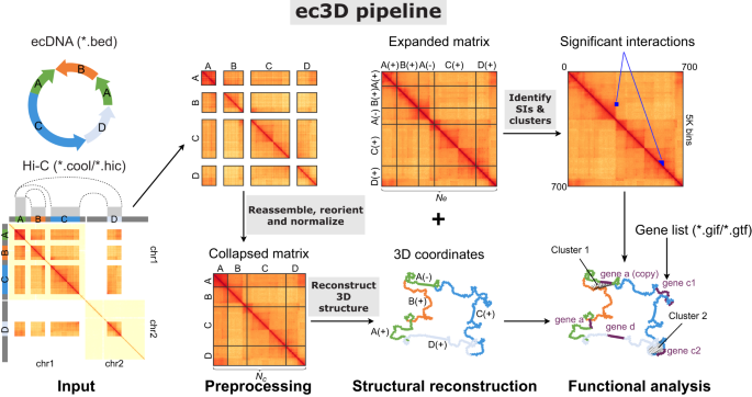
Chowdhury, Biswanath; Zhu, Kaiyuan; Li, Chaohui; Alsing, Jessica; Luebeck, Jens; Stefanova, Maria E; Chapman, Owen S; Kraft, Katerina; Zhang, Shu; Lim, Jun Yi Stanley; Xie, Yipeng; Kim, Yoon Jung; Wu, Sihan; Chavez, Lukas; Nir, Guy; Henssen, Anton G; Mischel, Paul S; Chang, Howard Y; Bafna, Vineet
Reconstructing the three-dimensional architecture of extrachromosomal DNA with ec3D Journal Article
In: Nat Commun, vol. 17, no. 1, pp. 894, 2025, ISSN: 2041-1723.
@article{pmid41422275,

title = {Reconstructing the three-dimensional architecture of extrachromosomal DNA with ec3D},

author = {Biswanath Chowdhury and Kaiyuan Zhu and Chaohui Li and Jessica Alsing and Jens Luebeck and Maria E Stefanova and Owen S Chapman and Katerina Kraft and Shu Zhang and Jun Yi Stanley Lim and Yipeng Xie and Yoon Jung Kim and Sihan Wu and Lukas Chavez and Guy Nir and Anton G Henssen and Paul S Mischel and Howard Y Chang and Vineet Bafna},

doi = {10.1038/s41467-025-67614-7},

issn = {2041-1723},

year  = {2025},

date = {2025-12-01},

journal = {Nat Commun},

volume = {17},

number = {1},

pages = {894},

abstract = {Extrachromosomal DNAs (ecDNAs) are circular DNA molecules prevalent in human cancers that drive tumor evolution and drug resistance. Their circular topology, which disrupts topological domains and rewires regulatory circuits, has typically been studied via pairwise interactions. Here we develop ec3D, a computational method for reconstructing three-dimensional ecDNA structures from Hi-C data. Given a candidate ecDNA sequence and whole-genome Hi-C data, ec3D reconstructs spatial structures by maximizing the Poisson likelihood of observed interactions. We validate ec3D using simulated structures, previously characterized cancer cell lines, and microscopy imaging. Our reconstructions reveal that ecDNAs occupy spherical configurations and mediate unique long-range regulatory interactions involved in gene regulation. Through algorithmic innovations, ec3D can resolve complex structures with duplicated segments, identify multi-way interactions, and identify potential intermolecular (trans) interactions. Our findings provide insights into how ecDNA's spatial organization bypasses normal chromosomal constraints and contributes to increased oncogene expression.},

keywords = {},

pubstate = {published},

tppubtype = {article}

}
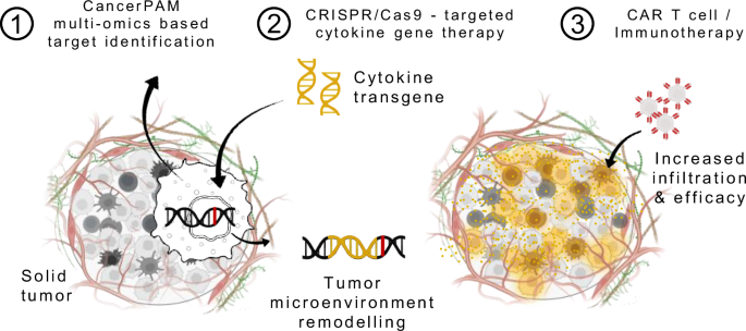
Launspach, Michael; Macos, Julia; Afzal, Shoaib; Hohmann, Janik; Appis, Marc L; Pilgram, Maximilian; Beez, Stefanie; Ohlendorf, Emily; van der Ven, Casper F T; Lachiheb, Chahrazad; Töws, Karin; Andersch, Lena; Jens, Marvin; Zirngibl, Felix; Kath, Jonas; Stecklum, Maria; Rodriguez-Fos, Elias; Anders, Kathleen; Wagner, Dimitrios L; Henssen, Anton G; Kühn, Ralf; Eggert, Angelika; Künkele, Annette
Personalized CRISPR knock-in cytokine gene therapy to remodel the tumor microenvironment and enhance CAR T cell therapy in solid tumors Journal Article
In: Nat Commun, vol. 16, no. 1, pp. 10987, 2025, ISSN: 2041-1723.
@article{pmid41366257,

title = {Personalized CRISPR knock-in cytokine gene therapy to remodel the tumor microenvironment and enhance CAR T cell therapy in solid tumors},

author = {Michael Launspach and Julia Macos and Shoaib Afzal and Janik Hohmann and Marc L Appis and Maximilian Pilgram and Stefanie Beez and Emily Ohlendorf and Casper F T van der Ven and Chahrazad Lachiheb and Karin Töws and Lena Andersch and Marvin Jens and Felix Zirngibl and Jonas Kath and Maria Stecklum and Elias Rodriguez-Fos and Kathleen Anders and Dimitrios L Wagner and Anton G Henssen and Ralf Kühn and Angelika Eggert and Annette Künkele},

doi = {10.1038/s41467-025-67328-w},

issn = {2041-1723},

year  = {2025},

date = {2025-12-01},

journal = {Nat Commun},

volume = {16},

number = {1},

pages = {10987},

abstract = {The immunosuppressive tumour microenvironment (TME) remains a central barrier to effective immunotherapy in solid tumours. We present a gene-therapeutic strategy that enables localized remodelling of the TME via tumour-intrinsic cytokine expression. Central to this approach is CancerPAM, a multi-omics bioinformatics pipeline that identifies and ranks patient-specific, tumour-exclusive CRISPR-Cas9 knock-in sites with high specificity and integration efficiency. Using neuroblastoma as a model, CancerPAM analysis of tumour sequencing data identifies optimal knock-in sites for pro-inflammatory cytokines (CXCL10, CXCL11, IFNG), and CancerPAM rankings correlate strongly with target-site specificity and knock-in efficiency, validating its predictive performance. CRISPR-mediated CXCL10 knock-in enhances CAR T cell infiltration and antitumour efficacy in vitro and in vivo, including humanized CD34⁺ HuNOG mice, where CXCL10-expressing tumours show stronger immune infiltration and prolonged tumour control within a reconstituted human immune microenvironment. Our findings establish a framework for safe and effective CRISPR-based cytokine delivery, integrating localized TME remodelling with cellular immunotherapies to enhance CAR T cells and other treatments in immune-refractory solid tumours.},

keywords = {},

pubstate = {published},

tppubtype = {article}

}
Montuori, Giulia; Tu, Fengyu; Qin, Di; Schmargon, Rachel; Rodriguez-Fos, Elias; Helmsauer, Konstantin; Hui, Hui; Mandal, Susmita; Purshouse, Karin; Fankhänel, Lara; Bosco, Bartolomeo; Spanjaard, Bastiaan; Seyboldt, Hannah; Grunewald, Laura; Schmitt, Matthias Jürgen; Gürgen, Dennis; Buck, Viktoria; Rosenfeldt, Mathias T; Dubois, Frank P B; Schallenberg, Simon; Lehmann, Annika; Theißen, Jessica; Taschner-Mandl, Sabine; Koch, Arend; Hundsdoerfer, Patrick; Künkele, Annette; Eggert, Angelika; Fischer, Matthias; Gargiulo, Gaetano; Krieger, Teresa G; Chavez, Lukas; Coscia, Fabian; Werner, Benjamin; Huang, Weini; Henssen, Anton G; Dörr, Jan R
Extrachromosomal DNA-Driven Oncogene Dosage Heterogeneity Promotes Rapid Adaptation to Therapy in MYCN-Amplified Cancers Journal Article
In: Cancer Discov, vol. 15, no. 10, pp. 2054–2077, 2025, ISSN: 2159-8290.
@article{pmid40773595,

title = {Extrachromosomal DNA-Driven Oncogene Dosage Heterogeneity Promotes Rapid Adaptation to Therapy in MYCN-Amplified Cancers},

author = {Giulia Montuori and Fengyu Tu and Di Qin and Rachel Schmargon and Elias Rodriguez-Fos and Konstantin Helmsauer and Hui Hui and Susmita Mandal and Karin Purshouse and Lara Fankhänel and Bartolomeo Bosco and Bastiaan Spanjaard and Hannah Seyboldt and Laura Grunewald and Matthias Jürgen Schmitt and Dennis Gürgen and Viktoria Buck and Mathias T Rosenfeldt and Frank P B Dubois and Simon Schallenberg and Annika Lehmann and Jessica Theißen and Sabine Taschner-Mandl and Arend Koch and Patrick Hundsdoerfer and Annette Künkele and Angelika Eggert and Matthias Fischer and Gaetano Gargiulo and Teresa G Krieger and Lukas Chavez and Fabian Coscia and Benjamin Werner and Weini Huang and Anton G Henssen and Jan R Dörr},

doi = {10.1158/2159-8290.CD-24-1738},

issn = {2159-8290},

year  = {2025},

date = {2025-10-01},

journal = {Cancer Discov},

volume = {15},

number = {10},

pages = {2054--2077},

abstract = {Extrachromosomal DNA (ecDNA) amplification enhances intercellular oncogene dosage variability and accelerates tumor evolution by violating foundational principles of genetic inheritance through its asymmetric mitotic segregation. Spotlighting high-risk neuroblastoma, we demonstrate how ecDNA amplification undermines the clinical efficacy of current therapies in cancers with extrachromosomal MYCN amplification. Integrating theoretical models of oncogene copy number-dependent fitness with single-cell ecDNA quantification and phenotype analyses, we reveal that ecDNA copy-number heterogeneity drives phenotypic diversity and determines treatment sensitivity through mechanisms unattainable by chromosomal oncogene amplification. We demonstrate that ecDNA copy number directly influences cell fate decisions in cancer cell lines, patient-derived xenografts, and primary neuroblastomas, illustrating how extrachromosomal oncogene dosage-driven phenotypic diversity offers a strong evolutionary advantage under therapeutic pressure. Furthermore, we identify senescent cells with reduced ecDNA copy numbers as a source of treatment resistance in neuroblastomas and outline a strategy for their targeted elimination to improve the treatment of MYCN-amplified cancers.nnSIGNIFICANCE: ecDNA-driven tumor genome evolution provides a major challenge to curative cancer therapies. We demonstrate that ecDNA copy-number dynamics drives treatment resistance by promoting oncogene dosage-dependent phenotypic heterogeneity in MYCN-amplified cancers. Exploiting phenotype-specific vulnerabilities of ecDNA cells, therefore, presents a powerful strategy to overcome treatment resistance. See related commentary by Korsah, p. 1979.},

keywords = {},

pubstate = {published},

tppubtype = {article}

}
Chan, Lap Kwan; Shan, Juanjuan; Rodriguez-Fos, Elias; Healy, Marc Eamonn; Leary, Peter; Parrotta, Rossella; Desboeufs, Nina; Semere, Gabriel; Wittstruck, Nadine; Henssen, Anton G; Weber, Achim
Extrachromosomal circular DNA promotes inflammation and hepatocellular carcinoma development Journal Article
In: Sci Adv, vol. 11, no. 42, pp. eadw0272, 2025, ISSN: 2375-2548.
@article{pmid41105787,

title = {Extrachromosomal circular DNA promotes inflammation and hepatocellular carcinoma development},

author = {Lap Kwan Chan and Juanjuan Shan and Elias Rodriguez-Fos and Marc Eamonn Healy and Peter Leary and Rossella Parrotta and Nina Desboeufs and Gabriel Semere and Nadine Wittstruck and Anton G Henssen and Achim Weber},

doi = {10.1126/sciadv.adw0272},

issn = {2375-2548},

year  = {2025},

date = {2025-10-01},

journal = {Sci Adv},

volume = {11},

number = {42},

pages = {eadw0272},

abstract = {Two decades after the initial report on increased micronuclei in human chronic liver disease (CLD) and hepatocellular carcinoma (HCC), their role in HCC development is still poorly understood. Here, we show that micronuclei in hepatocytes trigger a hepatic immune response and promote HCC development via an increased level of extrachromosomal circular DNA (eccDNA). Livers of a CLD model ( mice) show increased micronuclei and eccDNA levels. Circular sequencing confirms higher eccDNA levels in micronuclei compared to primary nuclei. The nuclei-segregated DNA fiber (NuSeF) assay we developed demonstrates that micronuclei are more susceptible to replication stress, exhibiting increased replication fork slowing. Comparing different murine liver disease models reveals that high eccDNA correlates with an increased tumor incidence. eccDNA is a strong immunostimulant and promotes a cross-talk between hepatocytes and immune cells through the cGAS-STING pathway. Deletion of  in  mice reduces immune cell chemotaxis and tumor incidence. Our findings suggest that eccDNA from micronuclei mediates inflammation-driven liver carcinogenesis in CLD.},

keywords = {},

pubstate = {published},

tppubtype = {article}

}

Sankar, Venkat; Hung, King L; Gnanasekar, Aditi; Wong, Ivy Tsz-Lo; Shi, Quanming; Kraft, Katerina; Jones, Matthew G; He, Britney Jiayu; Yan, Xiaowei; Belk, Julia A; Liu, Kevin J; Agarwal, Sangya; Wang, Sean K; Henssen, Anton G; Mischel, Paul S; Chang, Howard Y
Genetic elements promote retention of extrachromosomal DNA in cancer cells Journal Article
In: bioRxiv, 2025, ISSN: 2692-8205.
@article{pmid41278658,

title = {Genetic elements promote retention of extrachromosomal DNA in cancer cells},

author = {Venkat Sankar and King L Hung and Aditi Gnanasekar and Ivy Tsz-Lo Wong and Quanming Shi and Katerina Kraft and Matthew G Jones and Britney Jiayu He and Xiaowei Yan and Julia A Belk and Kevin J Liu and Sangya Agarwal and Sean K Wang and Anton G Henssen and Paul S Mischel and Howard Y Chang},

doi = {10.1101/2025.10.10.681495},

issn = {2692-8205},

year  = {2025},

date = {2025-10-01},

journal = {bioRxiv},

abstract = {Extrachromosomal DNA (ecDNA) is a prevalent and devastating form of oncogene amplification in cancer. Circular megabase-sized ecDNAs lack centromeres and segregate stochastically during cell division yet persist over many generations. EcDNAs were first observed to hitchhike on mitotic chromosomes into daughter cell nuclei over 40 years ago with unknown mechanism. Here we identify a family of human genomic elements, termed retention elements, that tether episomes to mitotic chromosomes to increase ecDNA transmission to daughter cells. We develop Retain-seq, a genome-scale assay that reveals thousands of human retention elements conferring generational persistence to heterologous episomes. Retention elements comprise a select set of CpG-rich gene promoters and act additively. Live-cell imaging and chromatin conformation capture show that retention elements physically interact with mitotic chromosomes at regions which are mitotically bookmarked by transcription factors and chromatin proteins, intermolecularly recapitulating promoter-enhancer interactions. Multiple retention elements are co-amplified with oncogenes on individual ecDNAs in human cancers and shape their sizes and structures. CpG-rich retention elements are focally hypomethylated; targeted cytosine methylation abrogates retention activity and leads to ecDNA loss, suggesting that methylation-sensitive interactions modulate episomal DNA retention. These results highlight the DNA elements and regulatory logic of mitotic ecDNA retention. Amplifications of retention elements promote the maintenance of oncogenic ecDNA across generations of cancer cells, revealing the principles of episome immortality intrinsic to the human genome.},

keywords = {},

pubstate = {published},

tppubtype = {article}

}
Chapman, Owen S; Sridhar, Sunita; Chow, Eugene Yui-Ching; Kenkre, Rishaan; Kirkland, Jonathan; Dutta, Aditi; Wang, Shanqing; Zhang, Wenshu; Brown, Miguel; Luebeck, Jens; Lo, Yan Yuen; Rodriguez-Fos, Elias; Henssen, Anton G; Okonechnikov, Konstantin; Ghasemi, David R; Pajtler, Kristian W; Kawauchi, Daisuke; Bafna, Vineet; Paul, Megan; Yip, Kevin; Mesirov, Jill P; Chavez, Lukas
Extrachromosomal DNA associates with poor survival across a broad spectrum of childhood solid tumors Journal Article
In: medRxiv, 2025.
@article{pmid40778132,

title = {Extrachromosomal DNA associates with poor survival across a broad spectrum of childhood solid tumors},

author = {Owen S Chapman and Sunita Sridhar and Eugene Yui-Ching Chow and Rishaan Kenkre and Jonathan Kirkland and Aditi Dutta and Shanqing Wang and Wenshu Zhang and Miguel Brown and Jens Luebeck and Yan Yuen Lo and Elias Rodriguez-Fos and Anton G Henssen and Konstantin Okonechnikov and David R Ghasemi and Kristian W Pajtler and Daisuke Kawauchi and Vineet Bafna and Megan Paul and Kevin Yip and Jill P Mesirov and Lukas Chavez},

doi = {10.1101/2025.07.22.24308163},

year  = {2025},

date = {2025-08-01},

journal = {medRxiv},

abstract = {Circular extrachromosomal DNA (ecDNA) is a common form of oncogene amplification in aggressive cancers. The frequency and diversity of ecDNA has been catalogued in adult and some childhood cancers; however, its role in most pediatric cancers is not well-understood. To address this gap, we accessed large pediatric cancer genomics data repositories and identified ecDNA from whole genome sequencing data using cloud computing. This retrospective cohort comprises 3,631 solid tumor biopsies from 2,968 patients covering all major childhood solid tumor types. Aggressive tumor types had particularly high incidences of ecDNA. Pediatric patients whose tumors harbored extrachromosomal DNA had significantly poorer five-year overall survival than children whose tumors contained only chromosomal amplifications. We catalogue known and potentially novel oncogenes recurrently amplified on ecDNA and show that ecDNA often evolves during disease progression. These results highlight patient populations that could potentially benefit from future ecDNA-directed therapies. To facilitate discovery, we developed an interactive catalogue of ecDNA in childhood cancer at https://ccdi-ecdna.org/.},

keywords = {},

pubstate = {published},

tppubtype = {article}

}
Yi, Eunhee; Gujar, Amit D; Zhao, Dacheng; Suina, Kentaro; Jin, Xue; Pardon, Katharina; Yu, Qinghao; Kagermazova, Larisa; Korsah, Emmanuel E; Dusseau, Noah A; Boeke, Jef D; Henssen, Anton G; Verhaak, Roel G W
Selective Depletion of Cancer Cells with Extrachromosomal DNA via Lentiviral Infection Journal Article
In: Cancer Res Commun, vol. 5, no. 8, pp. 1458–1465, 2025, ISSN: 2767-9764.
@article{pmid40787829,

title = {Selective Depletion of Cancer Cells with Extrachromosomal DNA via Lentiviral Infection},

author = {Eunhee Yi and Amit D Gujar and Dacheng Zhao and Kentaro Suina and Xue Jin and Katharina Pardon and Qinghao Yu and Larisa Kagermazova and Emmanuel E Korsah and Noah A Dusseau and Jef D Boeke and Anton G Henssen and Roel G W Verhaak},

doi = {10.1158/2767-9764.CRC-25-0144},

issn = {2767-9764},

year  = {2025},

date = {2025-08-01},

journal = {Cancer Res Commun},

volume = {5},

number = {8},

pages = {1458--1465},

abstract = {Extrachromosomal DNA (ecDNA), a major focal oncogene amplification mode found across cancer, has recently regained attention as an emerging cancer hallmark, with a pervasive presence across cancers. With technical advancements such as high-coverage sequencing and live-cell genome imaging, we can now investigate the behaviors and functions of ecDNA. However, we still lack an understanding of how to eliminate ecDNA. We observed depletion of cells containing ecDNA during lentiviral but not transposon-based transduction, whereas we sought to investigate the mechanism of ecDNA behavior. This discovery may provide critical information on utilizing a lentiviral system in emerging ecDNA research. Additionally, this observation suggests specific sensitivities for cells with ecDNA.nnSIGNIFICANCE: ecDNA is an essential factor in cancer progression. We found that a group of cancer cells with ecDNA is selectively depleted after lentiviral infection. This finding provides promise for ecDNA-specific targeting, suggests the need for caution in using lentivirus, and offers alternative ways to study ecDNA.},

keywords = {},

pubstate = {published},

tppubtype = {article}

}
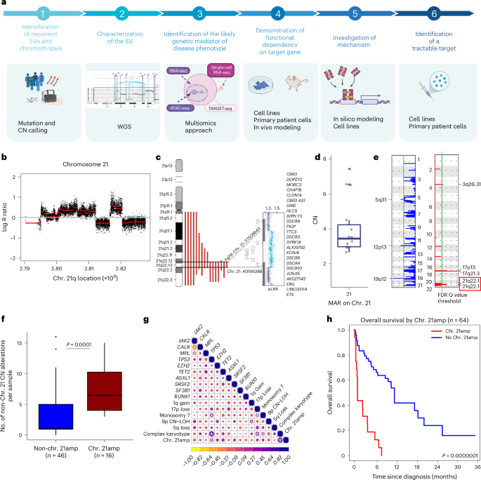
Brierley, Charlotte K; Yip, Bon Ham; Orlando, Giulia; Wen, Jeremy; Wen, Sean; Goyal, Harsh; Levine, Max; Jakobsdottir, G Maria; Tapinos, Avraam; Cornish, Alex J; Rodriguez-Romera, Antonio; Rodriguez-Meira, Alba; Bashton, Matthew; Hamblin, Angela; Clark, Sally Ann; Hamley, Joseph C; Fox, Olivia; Giurgiu, Madalina; O'Sullivan, Jennifer; Murphy, Lauren; Adamo, Assunta; Olijnik, Aude Anais; Cotton, Anitria; Hendrix, Emily; Narina, Shilpa; Pruett-Miller, Shondra M; Enshaei, Amir; Harrison, Claire; Drummond, Mark; Knapper, Steven; Tefferi, Ayalew; Antony-Debré, Iléana; Davies, James; Henssen, Anton G; Thongjuea, Supat; Wedge, David C; Constantinescu, Stefan N; Papaemmanuil, Elli; Psaila, Bethan; Crispino, John D; Mead, Adam J
Chromothripsis-associated chromosome 21 amplification orchestrates transformation to blast-phase MPN through targetable overexpression of DYRK1A Journal Article
In: Nat Genet, vol. 57, no. 6, pp. 1478–1492, 2025, ISSN: 1546-1718.
@article{pmid40490510,

title = {Chromothripsis-associated chromosome 21 amplification orchestrates transformation to blast-phase MPN through targetable overexpression of DYRK1A},

author = {Charlotte K Brierley and Bon Ham Yip and Giulia Orlando and Jeremy Wen and Sean Wen and Harsh Goyal and Max Levine and G Maria Jakobsdottir and Avraam Tapinos and Alex J Cornish and Antonio Rodriguez-Romera and Alba Rodriguez-Meira and Matthew Bashton and Angela Hamblin and Sally Ann Clark and Joseph C Hamley and Olivia Fox and Madalina Giurgiu and Jennifer O'Sullivan and Lauren Murphy and Assunta Adamo and Aude Anais Olijnik and Anitria Cotton and Emily Hendrix and Shilpa Narina and Shondra M Pruett-Miller and Amir Enshaei and Claire Harrison and Mark Drummond and Steven Knapper and Ayalew Tefferi and Iléana Antony-Debré and James Davies and Anton G Henssen and Supat Thongjuea and David C Wedge and Stefan N Constantinescu and Elli Papaemmanuil and Bethan Psaila and John D Crispino and Adam J Mead},

doi = {10.1038/s41588-025-02190-6},

issn = {1546-1718},

year  = {2025},

date = {2025-06-01},

journal = {Nat Genet},

volume = {57},

number = {6},

pages = {1478--1492},

abstract = {Chromothripsis, the chaotic shattering and repair of chromosomes, is common in cancer. Whether chromothripsis generates actionable therapeutic targets remains an open question. In a cohort of 64 patients in blast phase of a myeloproliferative neoplasm (BP-MPN), we describe recurrent amplification of a region of chromosome 21q ('chr. 21amp') in 25%, driven by chromothripsis in a third of these cases. We report that chr. 21amp BP-MPN has a particularly aggressive and treatment-resistant phenotype. DYRK1A, a serine threonine kinase, is the only gene in the 2.7-megabase minimally amplified region that showed both increased expression and chromatin accessibility compared with non-chr. 21amp BP-MPN controls. DYRK1A is a central node at the nexus of multiple cellular functions critical for BP-MPN development and is essential for BP-MPN cell proliferation in vitro and in vivo, and represents a druggable axis. Collectively, these findings define chr. 21amp as a prognostic biomarker in BP-MPN, and link chromothripsis to a therapeutic target.},

keywords = {},

pubstate = {published},

tppubtype = {article}

}
Rajkumar, Utkrisht; Prasad, Gino; Curtis, Ellis J; Wong, Ivy Tsz-Lo; Yan, Xiaowei; Zhang, Shu; Brückner, Lotte; Turner, Kristen; Wiese, Julie; Wahl, Justin; Wu, Sihan; Theissen, Jessica; Fischer, Matthias; Chang, Howard Y; Henssen, Anton G; Mischel, Paul S; Bafna, Vineet
Accurate Prediction of ecDNA in Interphase Cancer Cells using Deep Neural Networks Journal Article
In: bioRxiv, 2025, ISSN: 2692-8205.
@article{pmid40667255,

title = {Accurate Prediction of ecDNA in Interphase Cancer Cells using Deep Neural Networks},

author = {Utkrisht Rajkumar and Gino Prasad and Ellis J Curtis and Ivy Tsz-Lo Wong and Xiaowei Yan and Shu Zhang and Lotte Brückner and Kristen Turner and Julie Wiese and Justin Wahl and Sihan Wu and Jessica Theissen and Matthias Fischer and Howard Y Chang and Anton G Henssen and Paul S Mischel and Vineet Bafna},

doi = {10.1101/2025.06.23.661188},

issn = {2692-8205},

year  = {2025},

date = {2025-06-01},

journal = {bioRxiv},

abstract = {Oncogene amplification is a key driver of cancer pathogenesis and is often mediated by extrachromosomal DNA (ecDNA). EcDNA amplifications are associated with increased pathogenicity of cancer and poorer outcomes for patients. EcDNA can be detected accurately using fluorescence in situ hybridization (FISH) when cells are arrested in metaphase. However, the majority of cancer cells are non-mitotic and must be analyzed in interphase, where it is difficult to discern extrachromosomal amplifications from chromosomal amplifications. Thus, there is a need for methods that accurately predict oncogene amplification status from interphase cells. Here, we present interSeg, a deep learning-based tool to cytogenetically determine the amplification status as EC-amp, HSR-amp, or not amplified from interphase FISH images. We trained and validated interSeg on 652 images (40,446 nuclei). Tests on 215 cultured cell and tissue model images (9,733 nuclei) showed 89% and 97% accuracy at the nuclear and sample levels, respectively. The neuroblastoma patient tissue hold-out set (67 samples and 1,937 nuclei) also revealed 97% accuracy at the sample level in detecting the presence of focal amplification. In experimentally and computationally mixed images, interSeg accurately predicted the level of heterogeneity. The results showcase interSeg as an important method for analyzing oncogene amplifications.},

keywords = {},

pubstate = {published},

tppubtype = {article}

}
Rohde, Juliane; Henssen, Anton; Eggert, Angelika; Scheer, Monika
Rhabdomyosarcoma of head and neck varies in aggressiveness depending on the specific site of origin Journal Article
In: Oral Oncol, vol. 164, pp. 107263, 2025, ISSN: 1879-0593.
@article{pmid40188643,

title = {Rhabdomyosarcoma of head and neck varies in aggressiveness depending on the specific site of origin},

author = {Juliane Rohde and Anton Henssen and Angelika Eggert and Monika Scheer},

doi = {10.1016/j.oraloncology.2025.107263},

issn = {1879-0593},

year  = {2025},

date = {2025-05-01},

journal = {Oral Oncol},

volume = {164},

pages = {107263},

abstract = {OBJECTIVE: To evaluate predictive impact of granular subsites of head/neck rhabdomyosarcoma in a cross-age evaluation of the population-based SEER-program.nnDESIGN: Data were obtained for cases 0-90+ years, newly diagnosed with rhabdomyosarcoma at head/neck, registered in SEER17 2000-2020. Disease-specific survival (DSS) and overall survival (OS) were the endpoints, using the Kaplan-Meier estimator and Cox proportional hazards regression model. A granular site categorization was established.nnRESULTS: Median age of 1114 cases was 11 years. 5-year OS and DSS were 59.1 %±3.1 (95 %CI) and 62.4 %±3.1 with median follow-up for 662 survivors of 8.6 years. Increasing age was independently associated with worse prognosis. The rate of affected subsites varied considerably. Age, histology, tumor size, disease stage, the proportion of pathologically examined and affected lymph nodes differed significantly according to granular subsite. Granular subsites were of independent predictive impact when adjusted for age, size, histology, stage, and pathological lymph node status. While rhabdomyosarcoma at orbit, parotid gland, and ear correlated with best survival, larynx, oral cavity, paranasal sinuses, brain, pharynx, and nose were associated with adverse survival. In contrast to all other subsites, nasal and paranasal sinus rhabdomyosarcoma were predominantly alveolar, large, distant spread, and with the highest proportion of affected lymph nodes. Rhabdomyosarcoma of nose/paranasal sinuses exhibit high potential of spreading not only suggesting different biology but thorough staging including pathological lymph node assessment.nnCONCLUSION AND RELEVANCE: Granular head/neck subsites show different characteristics between subsites and highly varying outcomes. Understanding the impact of granular head/neck subsites on outcome may inform risk-adapted and novel approaches to rhabdomyosarcoma.},

keywords = {},

pubstate = {published},

tppubtype = {article}

}
Lv, Wei; Zeng, Yuchen; Li, Conghui; Liang, Yuan; Tao, Huiying; Zhu, Yanfen; Sui, Xiaolong; Li, Yue; Jiang, Shiqi; Gao, Qingqing; Rodriguez-Fos, Elias; Prasad, Gino; Wang, Yuanmei; Zhou, Run; Xu, Zhe; Pan, Xiaoguang; Chen, Linlin; Xiang, Xi; Teng, Huajing; Sun, Chaoyang; Qin, Tianyu; Dong, Wei; Li, Yongwei; Lan, Xun; Li, Xuesong; Lin, Lin; Bolund, Lars; Yang, Huanming; Verhaak, Roel G W; Faltas, Bishoy M; Hansen, Jacob B; Wu, Sihan; Mischel, Paul S; Henssen, Anton G; Bafna, Vineet; Luebeck, Jens; Regenberg, Birgitte; Luo, Yonglun; Lin, Chunhua; Han, Peng
Spatial-Temporal Diversity of Extrachromosomal DNA Shapes Urothelial Carcinoma Evolution and the Tumor Immune Microenvironment Journal Article
In: Cancer Discov, pp. OF1–OF22, 2025, ISSN: 2159-8290.
@article{pmid40331621,

title = {Spatial-Temporal Diversity of Extrachromosomal DNA Shapes Urothelial Carcinoma Evolution and the Tumor Immune Microenvironment},

author = {Wei Lv and Yuchen Zeng and Conghui Li and Yuan Liang and Huiying Tao and Yanfen Zhu and Xiaolong Sui and Yue Li and Shiqi Jiang and Qingqing Gao and Elias Rodriguez-Fos and Gino Prasad and Yuanmei Wang and Run Zhou and Zhe Xu and Xiaoguang Pan and Linlin Chen and Xi Xiang and Huajing Teng and Chaoyang Sun and Tianyu Qin and Wei Dong and Yongwei Li and Xun Lan and Xuesong Li and Lin Lin and Lars Bolund and Huanming Yang and Roel G W Verhaak and Bishoy M Faltas and Jacob B Hansen and Sihan Wu and Paul S Mischel and Anton G Henssen and Vineet Bafna and Jens Luebeck and Birgitte Regenberg and Yonglun Luo and Chunhua Lin and Peng Han},

doi = {10.1158/2159-8290.CD-24-1532},

issn = {2159-8290},

year  = {2025},

date = {2025-05-01},

journal = {Cancer Discov},

pages = {OF1--OF22},

abstract = {Our comprehensive analysis of ecDNA in urothelial carcinoma reveals its crucial role in driving the evolution and heterogeneity of multifocal cancer, as well as its early involvement in tumorigenesis. Moreover, this study sheds light on immune evasion mechanisms associated with ecDNA and offers valuable insights for developing targeted therapeutic strategies.},

keywords = {},

pubstate = {published},

tppubtype = {article}

}
Brückner, Lotte; Xu, Robin; Tang, Jun; Herrmann, Alexander; Wong, Ivy Tsz-Lo; Zhang, Shu; Tu, Fengyu; Pilon, Madison; Kukalev, Alexander; Pardon, Katharina; Sidorova, Olga; Atta, Joshua; Yu, Qinghao; Pradella, Davide; Ilić, Mila; Marco-Novais-Cruz,; Kaltenbach, Sarah; Treue, Denise; Giurgiu, Madalina; Herzog, Sergej; Hollinger, Ann-Sophie; Fernandez, Martina; Becker, Finnja; Louma, Varvara-Rigina; Schmargon, Rachel; Dörr, Jan; Gamlin, David; Lehmann, Annika; Gürgen, Dennis; Richter, Matthias; Dubois, Frank; Simeoni, Fabrizio; Pennycook, Betheney R; Hamilton, Alastair; Lindemann, Ralph K; Fischer, Matthias; Bafna, Vineet; Wahl, Geoffrey; Koche, Richard P; Chang, Howard Y; Papathanasiou, Stamatis; Medema, René; Spanjaard, Bastiaan; Ventura, Andrea; Pombo, Ana; Huang, Weini; Werner, Benjamin; Mischel, Paul S; Henssen, Anton G
Oncogene Silencing via ecDNA Micronucleation Journal Article
In: bioRxiv, 2025, ISSN: 2692-8205.
@article{pmid40321211,

title = {Oncogene Silencing via ecDNA Micronucleation},

author = {Lotte Brückner and Robin Xu and Jun Tang and Alexander Herrmann and Ivy Tsz-Lo Wong and Shu Zhang and Fengyu Tu and Madison Pilon and Alexander Kukalev and Katharina Pardon and Olga Sidorova and Joshua Atta and Qinghao Yu and Davide Pradella and Mila Ilić and Marco-Novais-Cruz and Sarah Kaltenbach and Denise Treue and Madalina Giurgiu and Sergej Herzog and Ann-Sophie Hollinger and Martina Fernandez and Finnja Becker and Varvara-Rigina Louma and Rachel Schmargon and Jan Dörr and David Gamlin and Annika Lehmann and Dennis Gürgen and Matthias Richter and Frank Dubois and Fabrizio Simeoni and Betheney R Pennycook and Alastair Hamilton and Ralph K Lindemann and Matthias Fischer and Vineet Bafna and Geoffrey Wahl and Richard P Koche and Howard Y Chang and Stamatis Papathanasiou and René Medema and Bastiaan Spanjaard and Andrea Ventura and Ana Pombo and Weini Huang and Benjamin Werner and Paul S Mischel and Anton G Henssen},

doi = {10.1101/2025.04.15.648906},

issn = {2692-8205},

year  = {2025},

date = {2025-04-01},

journal = {bioRxiv},

abstract = {Extrachromosomal DNA (ecDNA) is a common source of oncogene amplification across many types of cancer. The non-Mendelian inheritance of ecDNA contributes to heterogeneous tumour genomes that rapidly evolve to resist treatment. Here, using single-cell and live-cell imaging, single-micronucleus sequencing, and computational modelling, we demonstrate that elevated levels of ecDNA predisposes cells to micronucleation. Damage on ecDNA, commonly arising from replication stress, detaches ecDNA from the chromosomes upon which they hitchhike during cell division, thereby causing micronucleus formation in daughter cells. Clusters of oncogene-containing, CIP2A-TOPBP1-associated ecDNA molecules form, and asymmetrically segregate into daughter cell micronuclei during cell division. ecDNA chromatin remains highly active during mitosis, but upon micronucleation, it undergoes suppressive chromatin remodeling, largely ceasing oncogene transcription. These studies provide insight into the fate of damaged ecDNA during cell division.},

keywords = {},

pubstate = {published},

tppubtype = {article}

}
Lv, Wei; Zeng, Yuchen; Li, Conghui; Liang, Yuan; Tao, Huiying; Zhu, Yanfen; Sui, Xiaolong; Li, Yue; Jiang, Shiqi; Gao, Qingqing; Rodriguez-Fos, Elias; Prasad, Gino; Wang, Yuanmei; Zhou, Run; Xu, Zhe; Pan, Xiaoguang; Chen, Linlin; Xiang, Xi; Teng, Huajing; Sun, Chaoyang; Qin, Tianyu; Dong, Wei; Li, Yongwei; Lan, Xun; Li, Xuesong; Lin, Lin; Bolund, Lars; Yang, Huanming; Verhaak, Roel G W; Faltas, Bishoy M; Hansen, Jacob B; Wu, Sihan; Mischel, Paul S; Henssen, Anton G; Bafna, Vineet; Luebeck, Jens; Regenberg, Birgitte; Luo, Yonglun; Lin, Chunhua; Han, Peng
Spatial-Temporal Diversity of Extrachromosomal DNA Shapes Urothelial Carcinoma Evolution and Tumor-Immune Microenvironment Journal Article
In: Cancer Discov, 2025, ISSN: 2159-8290.
@article{pmid40062518,

title = {Spatial-Temporal Diversity of Extrachromosomal DNA Shapes Urothelial Carcinoma Evolution and Tumor-Immune Microenvironment},

author = {Wei Lv and Yuchen Zeng and Conghui Li and Yuan Liang and Huiying Tao and Yanfen Zhu and Xiaolong Sui and Yue Li and Shiqi Jiang and Qingqing Gao and Elias Rodriguez-Fos and Gino Prasad and Yuanmei Wang and Run Zhou and Zhe Xu and Xiaoguang Pan and Linlin Chen and Xi Xiang and Huajing Teng and Chaoyang Sun and Tianyu Qin and Wei Dong and Yongwei Li and Xun Lan and Xuesong Li and Lin Lin and Lars Bolund and Huanming Yang and Roel G W Verhaak and Bishoy M Faltas and Jacob B Hansen and Sihan Wu and Paul S Mischel and Anton G Henssen and Vineet Bafna and Jens Luebeck and Birgitte Regenberg and Yonglun Luo and Chunhua Lin and Peng Han},

doi = {10.1158/2159-8290.CD-24-1532},

issn = {2159-8290},

year  = {2025},

date = {2025-03-01},

journal = {Cancer Discov},

abstract = {Extrachromosomal DNA (ecDNA) presents a promising target for cancer therapy; however, its spatial-temporal diversity and influence on tumor evolution and the immune microenvironment remain largely unclear. We apply computational methods to analyze ecDNA from whole-genome sequencing data of 595 urothelial carcinoma (UC) patients. We demonstrate that ecDNA drives clonal evolution through structural rearrangements during malignant transformation and recurrence of UC. This supports a model wherein tumors evolve via the selective expansion of ecDNA-bearing cells. Through multi-regional sampling of tumors, we demonstrate that ecDNA contributes to the evolution of multifocality and increased intratumoral heterogeneity. EcDNA is present in 36% of UC tumors and correlates with an immunosuppressive phenotype and poor prognosis. Single-cell RNA sequencing analyses reveal that ecDNA+ malignant cells exhibit diminished expression of major histocompatibility complex class I molecules, enabling them to evade T-cell immunity. Finally, we show that sequencing of urinary sediment-derived DNA has excellent specificity in detecting ecDNA.},

keywords = {},

pubstate = {published},

tppubtype = {article}

}
Yi, Eunhee; Gujar, Amit D; Zhao, Dacheng; Suina, Kentaro; Jin, Xue; Pardon, Katharina; Yu, Qinghao; Kagermazova, Larisa; Boeke, Jef D; Henssen, Anton G; Verhaak, Roel G W
Selective depletion of cancer cells with extrachromosomal DNA via lentiviral infection Journal Article
In: bioRxiv, 2025, ISSN: 2692-8205.
@article{pmid40060579,

title = {Selective depletion of cancer cells with extrachromosomal DNA via lentiviral infection},

author = {Eunhee Yi and Amit D Gujar and Dacheng Zhao and Kentaro Suina and Xue Jin and Katharina Pardon and Qinghao Yu and Larisa Kagermazova and Jef D Boeke and Anton G Henssen and Roel G W Verhaak},

doi = {10.1101/2025.02.22.639631},

issn = {2692-8205},

year  = {2025},

date = {2025-02-01},

journal = {bioRxiv},

abstract = {Dear Editor Extrachromosomal DNA (ecDNA), a major focal oncogene amplification mode found across cancer, has recently regained attention as an emerging cancer hallmark  , with a pervasive presence across cancers  . With technical advancements such as high-coverage sequencing and live-cell genome imaging, we can now investigate ecDNA's behaviors and functions  . However, we still lack an understanding of how to eliminate ecDNA. We observed depletion of cells containing ecDNA during lentiviral but not transposon-based transduction while we sought to investigate the mechanism of ecDNA behavior. This discovery may provide critical information on utilizing a lentiviral system in emerging ecDNA research. Additionally, this observation suggests specific sensitivities for cells with ecDNA.},

keywords = {},

pubstate = {published},

tppubtype = {article}

}
Grunewald, Laura; Andersch, Lena; Helmsauer, Konstantin; Schwiebert, Silke; Klaus, Anika; Henssen, Anton G; Straka, Teresa; Lodrini, Marco; Wicha, Sebastian G; Fuchs, Steffen; Hertwig, Falk; Westermann, Frank; Vitali, Alice; Caramel, Carlotta; Büchel, Gabriele; Eilers, Martin; Astrahantseff, Kathy; Eggert, Angelika; Höpken, Uta E; Schulte, Johannes H; Blankenstein, Thomas; Anders, Kathleen; Künkele, Annette
Targeting MYCN upregulates L1CAM tumor antigen in MYCN-dysregulated neuroblastoma to increase CAR T cell efficacy Journal Article
In: Pharmacol Res, vol. 212, pp. 107608, 2025, ISSN: 1096-1186.
@article{pmid39828101,

title = {Targeting MYCN upregulates L1CAM tumor antigen in MYCN-dysregulated neuroblastoma to increase CAR T cell efficacy},

author = {Laura Grunewald and Lena Andersch and Konstantin Helmsauer and Silke Schwiebert and Anika Klaus and Anton G Henssen and Teresa Straka and Marco Lodrini and Sebastian G Wicha and Steffen Fuchs and Falk Hertwig and Frank Westermann and Alice Vitali and Carlotta Caramel and Gabriele Büchel and Martin Eilers and Kathy Astrahantseff and Angelika Eggert and Uta E Höpken and Johannes H Schulte and Thomas Blankenstein and Kathleen Anders and Annette Künkele},

doi = {10.1016/j.phrs.2025.107608},

issn = {1096-1186},

year  = {2025},

date = {2025-02-01},

journal = {Pharmacol Res},

volume = {212},

pages = {107608},

abstract = {Current treatment protocols have limited success against MYCN-amplified neuroblastoma. Adoptive T cell therapy presents an innovative strategy to improve cure rates. However, L1CAM-targeting CAR T cells achieved only limited response against refractory/relapsed neuroblastoma so far. We investigated how oncogenic MYCN levels influence tumor cell response to CAR T cells, as one possible factor limiting clinical success. A MYCN-inducible neuroblastoma cell model was created. L1CAM-CAR T cell effector function was assessed (activation markers, cytokine release, tumor cytotoxicity) after coculture with the model or MYCN-amplified neuroblastoma cell lines. RNA sequencing datasets characterizing the model were compared to publicly available RNA/proteomic datasets. MYCN-directed L1CAM regulation was explored using public ChIP-sequencing datasets. Synergism between CAR T cells and the indirect MYCN inhibitor, MLN8237, was assessed in vitro using the Bliss model and in vivo in an immunocompromised mouse model. Inducing high MYCN levels in the neuroblastoma cell model reduced L1CAM expression and, consequently, L1CAM-CAR T cell effector function in vitro. Primary neuroblastomas possessing high MYCN levels expressed lower levels of both the L1CAM transcript and L1CAM tumor antigen. MLN8237 treatment restored L1CAM tumor expression and L1CAM-CAR T cell effector function. Combining MLN8237 and L1CAM-CAR T cell treatment synergistically enhanced MYCN-overexpressing tumor cytotoxicity in vitro and in vivo concomitant with severe in vivo toxicity. We identify target antigen downregulation as source of resistance against L1CAM-CAR T cells in MYCN-driven neuroblastoma cells. These data suggest that L1CAM-CAR T cell therapy combined with pharmacological MYCN inhibition may benefit patients with MYCN-amplified neuroblastoma.},

keywords = {},

pubstate = {published},

tppubtype = {article}

}

Andersch, Lena; Grunewald, Laura; Stecklum, Maria; Klironomos, Filippos; Haase, Kerstin; Hollek, Viola; Lam, Tobias; Jung, Beate Anahita; Winkler, Anika; Schwiebert, Silke; Astrahantseff, Kathy; Launspach, Michael; Jens, Marvin; Henssen, Anton; Kloke, Lutz; Blüthgen, Nils; Eggert, Angelika; Schulte, Johannes H; Anders, Kathleen; Künkele, Annette
Investigating genetic modifications to enhance L1CAM-CAR T cell migration in solid tumors in a 3D bioprinted neuroblastoma model Journal Article
In: Front Immunol, vol. 16, pp. 1677361, 2025, ISSN: 1664-3224.
@article{pmid41394860,

title = {Investigating genetic modifications to enhance L1CAM-CAR T cell migration in solid tumors in a 3D bioprinted neuroblastoma model},

author = {Lena Andersch and Laura Grunewald and Maria Stecklum and Filippos Klironomos and Kerstin Haase and Viola Hollek and Tobias Lam and Beate Anahita Jung and Anika Winkler and Silke Schwiebert and Kathy Astrahantseff and Michael Launspach and Marvin Jens and Anton Henssen and Lutz Kloke and Nils Blüthgen and Angelika Eggert and Johannes H Schulte and Kathleen Anders and Annette Künkele},

doi = {10.3389/fimmu.2025.1677361},

issn = {1664-3224},

year  = {2025},

date = {2025-01-01},

journal = {Front Immunol},

volume = {16},

pages = {1677361},

abstract = {INTRODUCTION: Effective CAR T cell infiltration into solid tumors remains a major barrier to therapy success. Despite their clinical potential, few studies have evaluated phenotypes of CAR T cells successfully invading the tumor mass following infusion. Phenotypic information would enrich our understanding of the mechanisms governing CAR T cell migration into solid tumors. Here we implemented an  strategy to identify genes driving L1CAM-CAR T cell migration into a 3D tumor mass.nnMETHODS: L1CAM-CAR T cells were separated into 2 groups by their capability to infiltrate (or not) a 3D bioprinted neuroblastoma model. Single-cell and bulk RNA sequencing was performed, and infiltrating CAR T cells were compared to noninfiltrating cells to seek genetic drivers of CAR T cell migration. CRISPR/Cas9 technology was used to generate modified L1CAM-CAR T cells.nnRESULTS: Tumor-infiltrating L1CAM-CAR T cells expressed lower levels of the selectin P ligand (SELPLG) glycoprotein and higher levels of the T cell-specific adaptor protein, SH2D2A. Functional characterization of L1CAM-CAR T cells genetically modified to enforce these characteristics demonstrated that neither trait negatively impacted L1CAM-CAR T cell cytotoxicity, activation and cytokine release upon coculture with neuroblastoma target cells. Transgenic SH2D2A expression did not improve CAR T cell migration in an endothelial transmembrane assay. SELPLG knockout benefited CAR T cell  trans-endothelial migration, but did not enhance anti-tumor efficacy in an immunodeficient mouse model.nnDISCUSSION: Our findings reveal a key limitation of murine xenograft models, which are widely used as the gold standard for preclinical CAR T cell testing. The lack of conservation between the human and murine SELPLG proteins likely accounts for the discrepancy between enhanced  migration of SELPLG-deficient L1CAM-CAR T cells and their lack of improved efficacy in the mouse model. This underscores the need for more predictive human-relevant models to better preclinically evaluate CAR T cell function.},

keywords = {},

pubstate = {published},

tppubtype = {article}

}
Hollander, Jan F; Szymansky, Annabell; Wünschel, Jasmin; Astrahantseff, Kathy; Rosswog, Carolina; Thorwarth, Anne; Thole-Kliesch, Theresa M; González, Rocío Chamorro; Hundsdörfer, Patrick; Hauptmann, Kathrin; Schmelz, Karin; Gürgen, Dennis; Rogasch, Julian M M; Henssen, Anton G; Fischer, Matthias; Schulte, Johannes H; Eckert, Cornelia; Eggert, Angelika; Lodrini, Marco; Deubzer, Hedwig E
Serially Quantifying TERT Rearrangement Breakpoints in ctDNA Enables Minimal Residual Disease Monitoring in Patients with Neuroblastoma Journal Article
In: Cancer Res Commun, vol. 5, no. 1, pp. 167–177, 2025, ISSN: 2767-9764.
@article{pmid39760332,

title = {Serially Quantifying TERT Rearrangement Breakpoints in ctDNA Enables Minimal Residual Disease Monitoring in Patients with Neuroblastoma},

author = {Jan F Hollander and Annabell Szymansky and Jasmin Wünschel and Kathy Astrahantseff and Carolina Rosswog and Anne Thorwarth and Theresa M Thole-Kliesch and Rocío Chamorro González and Patrick Hundsdörfer and Kathrin Hauptmann and Karin Schmelz and Dennis Gürgen and Julian M M Rogasch and Anton G Henssen and Matthias Fischer and Johannes H Schulte and Cornelia Eckert and Angelika Eggert and Marco Lodrini and Hedwig E Deubzer},

doi = {10.1158/2767-9764.CRC-24-0569},

issn = {2767-9764},

year  = {2025},

date = {2025-01-01},

journal = {Cancer Res Commun},

volume = {5},

number = {1},

pages = {167--177},

abstract = {ABSTRACT: Telomerase is reactivated by genomic TERT rearrangements in ∼30% of diagnosed high-risk neuroblastomas. Dismal patient prognosis results if the RAS/MAPK/ALK signaling transduction network also harbors mutations. We present a liquid biopsy–based monitoring strategy for this particularly vulnerable pediatric patient subgroup for which real-time molecular diagnostic tools are limited to date. Droplet digital PCR assays quantifying patient-individualized TERT rearrangement breakpoint copies, ALK copy numbers, and allelic ALK p.R1275Q mutation frequencies were applied to longitudinally collected liquid biopsies (peripheral blood and bone marrow [BM] plasma, n = 44 biosamples), the mononuclear cell fraction from BM and matched tumor samples. Marker detection was compared with current gold-standard diagnostics. Reanalysis of whole-genome and targeted panel sequencing data from 169 patients identified 64 TERT-rearranged neuroblastoma samples collected at initial and/or relapse diagnosis from 55 patients (254 total TERT rearrangement events). Detection and quantification of unique TERT rearrangement breakpoints in as little as 1 ng of total cell-free DNA in peripheral blood plasma improved therapy response assessment and early relapse detection in individual patients. Proof-of-concept is provided for minimal residual disease detection in the BM niche, from which relapses frequently arise, by analyzing unique TERT rearrangement breakpoints in BM plasma–derived cell-free DNA. TERT rearrangement breakpoints, as a single marker or combined with mutations in the RAS/MAPK/ALK signaling transduction network, can serve as robust and highly sensitive biomarkers for disease activity and spatially and temporally resolve disease better than current gold-standard diagnostics in individual patients with TERT-driven neuroblastoma.nnSIGNIFICANCE: Real-time molecular monitoring of TERT-rearranged high-risk neuroblastoma is an unmet clinical need. We tested liquid biopsy-based assays for patient-individualized TERT breakpoint sequences to monitor disease in pediatric patients. Our digital PCR approach provides high resolution of spatial and temporal disease quantification in individual patients and is applicable for clinical routine.},

keywords = {},

pubstate = {published},

tppubtype = {article}

}
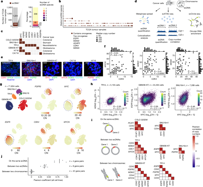
Hung, King L; Jones, Matthew G; Wong, Ivy Tsz-Lo; Curtis, Ellis J; Lange, Joshua T; He, Britney Jiayu; Luebeck, Jens; Schmargon, Rachel; Scanu, Elisa; Brückner, Lotte; Yan, Xiaowei; Li, Rui; Gnanasekar, Aditi; González, Rocío Chamorro; Belk, Julia A; Liu, Zhonglin; Melillo, Bruno; Bafna, Vineet; Dörr, Jan R; Werner, Benjamin; Huang, Weini; Cravatt, Benjamin F; Henssen, Anton G; Mischel, Paul S; Chang, Howard Y
Coordinated inheritance of extrachromosomal DNAs in cancer cells Journal Article
In: Nature, vol. 635, no. 8037, pp. 201–209, 2024, ISSN: 1476-4687.
@article{pmid39506152,

title = {Coordinated inheritance of extrachromosomal DNAs in cancer cells},

author = {King L Hung and Matthew G Jones and Ivy Tsz-Lo Wong and Ellis J Curtis and Joshua T Lange and Britney Jiayu He and Jens Luebeck and Rachel Schmargon and Elisa Scanu and Lotte Brückner and Xiaowei Yan and Rui Li and Aditi Gnanasekar and Rocío Chamorro González and Julia A Belk and Zhonglin Liu and Bruno Melillo and Vineet Bafna and Jan R Dörr and Benjamin Werner and Weini Huang and Benjamin F Cravatt and Anton G Henssen and Paul S Mischel and Howard Y Chang},

doi = {10.1038/s41586-024-07861-8},

issn = {1476-4687},

year  = {2024},

date = {2024-11-01},

journal = {Nature},

volume = {635},

number = {8037},

pages = {201--209},

abstract = {The chromosomal theory of inheritance dictates that genes on the same chromosome segregate together while genes on different chromosomes assort independently. Extrachromosomal DNAs (ecDNAs) are common in cancer and drive oncogene amplification, dysregulated gene expression and intratumoural heterogeneity through random segregation during cell division. Distinct ecDNA sequences, termed ecDNA species, can co-exist to facilitate intermolecular cooperation in cancer cells. How multiple ecDNA species within a tumour cell are assorted and maintained across somatic cell generations is unclear. Here we show that cooperative ecDNA species are coordinately inherited through mitotic co-segregation. Imaging and single-cell analyses show that multiple ecDNAs encoding distinct oncogenes co-occur and are correlated in copy number in human cancer cells. ecDNA species are coordinately segregated asymmetrically during mitosis, resulting in daughter cells with simultaneous copy-number gains in multiple ecDNA species before any selection. Intermolecular proximity and active transcription at the start of mitosis facilitate the coordinated segregation of ecDNA species, and transcription inhibition reduces co-segregation. Computational modelling reveals the quantitative principles of ecDNA co-segregation and co-selection, predicting their observed distributions in cancer cells. Coordinated inheritance of ecDNAs enables co-amplification of specialized ecDNAs containing only enhancer elements and guides therapeutic strategies to jointly deplete cooperating ecDNA oncogenes. Coordinated inheritance of ecDNAs confers stability to oncogene cooperation and novel gene regulatory circuits, allowing winning combinations of epigenetic states to be transmitted across cell generations.},

keywords = {},

pubstate = {published},

tppubtype = {article}

}
Kern, Leonie; Henssen, Anton; Eggert, Angelika; Scheer, Monika
The Other Site of Rhabdomyosarcoma Journal Article
In: Cancer Med, vol. 13, no. 20, pp. e70348, 2024, ISSN: 2045-7634.
@article{pmid39463067,

title = {The Other Site of Rhabdomyosarcoma},

author = {Leonie Kern and Anton Henssen and Angelika Eggert and Monika Scheer},

doi = {10.1002/cam4.70348},

issn = {2045-7634},

year  = {2024},

date = {2024-10-01},

journal = {Cancer Med},

volume = {13},

number = {20},

pages = {e70348},

abstract = {BACKGROUND: Rhabdomyosarcoma (RMS) is a rare malignant soft tissue sarcoma (STS), accounting for almost 50% of pediatric STSs. Due to its heterogeneity, RMS presents challenges in diagnosis and treatment, with prognosis varying depending on multiple factors. Tumors localized in the other site (OTH)-including the paraspinal, perianal, thoracic, abdominal, pelvic, and perineal regions-are generally classified as unfavorable. This study assesses the clinical features and prognoses of RMS in OTH locations depending on its site of origin.nnMETHODS: An explorative analysis of RMS cases from the SEER 17 database 2000-2020 was conducted. Patients of all ages with histologically confirmed RMS as primary malignant disease classified under OTH, were included. OTH was categorized in four granular site classifications. Overall survival (OS) and disease-specific survival (DSS) were analyzed using Kaplan-Meier estimators. Factors independently influencing survival, including a site classification model presented in this study, were identified through Cox regression analysis.nnRESULTS: Out of 4168 patients with RMS, 990 cases of RMS with the OTH site met the inclusion criteria. The median age was 16 years. The predominant histological subtypes were embryonal (33.0%) and alveolar (25.5%). Most tumors were ≥ 5 cm (median 9 cm) and located primarily in the pelvic region (41.5%). The 3-, 5-, and 10-year OS rates were 45.4% ± 3.332 (95% CI), 40.7 ± 3.332, and 38.6% ± 3.332, respectively, while DSS rates were 43.3% ± 3.136 (95% CI), 38.3% ± 3.136, and 35.1% ± 3.332. In the multivariate analysis age, histological type, site in a granular categorization, stage, regional lymph node examination, and regional lymph node involvement (pathologically proven) were independently associated with survival. Through both univariate and multivariate analyses, an OTH favorable group could be established. The OTH favorable group consists of the anal region, gallbladder and biliary tract, and breast.nnCONCLUSION: RMS in OTH shows significant differences in prognosis, putting the current categorization as unfavorable into question and making a more detailed classification necessary. Furthermore, pathological regional lymph node assessment is specifically in the OTH localization recommended.},

keywords = {},

pubstate = {published},

tppubtype = {article}

}
Giurgiu, Mădălina; Wittstruck, Nadine; Rodriguez-Fos, Elias; González, Rocío Chamorro; Brückner, Lotte; Krienelke-Szymansky, Annabell; Helmsauer, Konstantin; Hartebrodt, Anne; Euskirchen, Philipp; Koche, Richard P; Haase, Kerstin; Reinert, Knut; Henssen, Anton G
Reconstructing extrachromosomal DNA structural heterogeneity from long-read sequencing data using Decoil Journal Article
In: Genome Res, vol. 34, no. 9, pp. 1355–1364, 2024, ISSN: 1549-5469.
@article{pmid39111816,

title = {Reconstructing extrachromosomal DNA structural heterogeneity from long-read sequencing data using Decoil},

author = {Mădălina Giurgiu and Nadine Wittstruck and Elias Rodriguez-Fos and Rocío Chamorro González and Lotte Brückner and Annabell Krienelke-Szymansky and Konstantin Helmsauer and Anne Hartebrodt and Philipp Euskirchen and Richard P Koche and Kerstin Haase and Knut Reinert and Anton G Henssen},

doi = {10.1101/gr.279123.124},

issn = {1549-5469},

year  = {2024},

date = {2024-10-01},

journal = {Genome Res},

volume = {34},

number = {9},

pages = {1355--1364},

abstract = {Circular extrachromosomal DNA (ecDNA) is a form of oncogene amplification found across cancer types and associated with poor outcome in patients. ecDNA can be structurally complex and can contain rearranged DNA sequences derived from multiple chromosome locations. As the structure of ecDNA can impact oncogene regulation and may indicate mechanisms of its formation, disentangling it at high resolution from sequencing data is essential. Even though methods have been developed to identify and reconstruct ecDNA in cancer genome sequencing, it remains challenging to resolve complex ecDNA structures, in particular amplicons with shared genomic footprints. We here introduce Decoil, a computational method that combines a breakpoint-graph approach with  regression to reconstruct complex ecDNA and deconvolve co-occurring ecDNA elements with overlapping genomic footprints from long-read nanopore sequencing. Decoil outperforms de novo assembly and alignment-based methods in simulated long-read sequencing data for both simple and complex ecDNAs. Applying Decoil on whole-genome sequencing data uncovered different ecDNA topologies and explored ecDNA structure heterogeneity in neuroblastoma tumors and cell lines, indicating that this method may improve ecDNA structural analyses in cancer.},

keywords = {},

pubstate = {published},

tppubtype = {article}

}
Stöber, Maja C; González, Rocío Chamorro; Brückner, Lotte; Conrad, Thomas; Wittstruck, Nadine; Szymansky, Annabell; Eggert, Angelika; Schulte, Johannes H; Koche, Richard P; Henssen, Anton G; Schwarz, Roland F; Haase, Kerstin
Intercellular extrachromosomal DNA copy-number heterogeneity drives neuroblastoma cell state diversity Journal Article
In: Cell Rep, vol. 43, no. 9, pp. 114711, 2024, ISSN: 2211-1247.
@article{pmid39255063,

title = {Intercellular extrachromosomal DNA copy-number heterogeneity drives neuroblastoma cell state diversity},

author = {Maja C Stöber and Rocío Chamorro González and Lotte Brückner and Thomas Conrad and Nadine Wittstruck and Annabell Szymansky and Angelika Eggert and Johannes H Schulte and Richard P Koche and Anton G Henssen and Roland F Schwarz and Kerstin Haase},

doi = {10.1016/j.celrep.2024.114711},

issn = {2211-1247},

year  = {2024},

date = {2024-09-01},

journal = {Cell Rep},

volume = {43},

number = {9},

pages = {114711},

abstract = {Neuroblastoma exhibits significant inter- and intra-tumor genetic heterogeneity and varying clinical outcomes. Extrachromosomal DNAs (ecDNAs) may drive this heterogeneity by independently segregating during cell division, leading to rapid oncogene amplification. While ecDNA-mediated oncogene amplification is linked to poor prognosis in various cancers, the effects of ecDNA copy-number heterogeneity on intermediate phenotypes are poorly understood. Here, we leverage DNA and RNA sequencing from the same single cells in cell lines and neuroblastoma patients to investigate these effects. By analyzing ecDNA amplicon structures, we reveal extensive intercellular ecDNA copy-number heterogeneity. We also provide direct evidence of how this heterogeneity influences the expression of cargo genes, including MYCN and its downstream targets, and the overall transcriptional state of neuroblastoma cells. Our findings highlight the role of ecDNA copy number in promoting rapid adaptability of cellular states within tumors, underscoring the need for ecDNA-specific treatment strategies to address tumor formation and adaptation.},

keywords = {},

pubstate = {published},

tppubtype = {article}

}
Pusch, Fabian F; García, Heathcliff Dorado; Xu, Robin; Gürgen, Dennis; Bei, Yi; Brückner, Lotte; Röefzaad, Claudia; von Stebut, Jennifer; Bardinet, Victor; Gonzalez, Rocío Chamorro; Eggert, Angelika; Schulte, Johannes H; Hundsdörfer, Patrick; Seifert, Georg; Haase, Kerstin; Schäfer, Beat W; Wachtel, Marco; Kühl, Anja A; Ortiz, Michael V; Wengner, Antje M; Scheer, Monika; Henssen, Anton G
Elimusertib has Antitumor Activity in Preclinical Patient-Derived Pediatric Solid Tumor Models Journal Article
In: Mol Cancer Ther, vol. 23, no. 4, pp. 507–519, 2024, ISSN: 1538-8514.
@article{pmid38159110,

title = {Elimusertib has Antitumor Activity in Preclinical Patient-Derived Pediatric Solid Tumor Models},

author = {Fabian F Pusch and Heathcliff Dorado García and Robin Xu and Dennis Gürgen and Yi Bei and Lotte Brückner and Claudia Röefzaad and Jennifer von Stebut and Victor Bardinet and Rocío Chamorro Gonzalez and Angelika Eggert and Johannes H Schulte and Patrick Hundsdörfer and Georg Seifert and Kerstin Haase and Beat W Schäfer and Marco Wachtel and Anja A Kühl and Michael V Ortiz and Antje M Wengner and Monika Scheer and Anton G Henssen},

doi = {10.1158/1535-7163.MCT-23-0094},

issn = {1538-8514},

year  = {2024},

date = {2024-04-01},

journal = {Mol Cancer Ther},

volume = {23},

number = {4},

pages = {507--519},

abstract = {The small-molecule inhibitor of ataxia telangiectasia and Rad3-related protein (ATR), elimusertib, is currently being tested clinically in various cancer entities in adults and children. Its preclinical antitumor activity in pediatric malignancies, however, is largely unknown. We here assessed the preclinical activity of elimusertib in 38 cell lines and 32 patient-derived xenograft (PDX) models derived from common pediatric solid tumor entities. Detailed in vitro and in vivo molecular characterization of the treated models enabled the evaluation of response biomarkers. Pronounced objective response rates were observed for elimusertib monotherapy in PDX, when treated with a regimen currently used in clinical trials. Strikingly, elimusertib showed stronger antitumor effects than some standard-of-care chemotherapies, particularly in alveolar rhabdomysarcoma PDX. Thus, elimusertib has strong preclinical antitumor activity in pediatric solid tumor models, which may translate to clinically meaningful responses in patients.},

keywords = {},

pubstate = {published},

tppubtype = {article}

}
Bei, Yi; Bramé, Luca; Kirchner, Marieluise; Fritsche-Guenther, Raphaela; Kunz, Severine; Bhattacharya, Animesh; Rusu, Mara-Camelia; Gürgen, Dennis; Dubios, Frank P B; Köppke, Julia K C; Proba, Jutta; Wittstruck, Nadine; Sidorova, Olga Alexandra; González, Rocío Chamorro; Garcia, Heathcliff Dorado; Brückner, Lotte; Xu, Robin; Giurgiu, Mădălina; Rodriguez-Fos, Elias; Yu, Qinghao; Spanjaard, Bastiaan; Koche, Richard P; Schmitt, Clemens A; Schulte, Johannes H; Eggert, Angelika; Haase, Kerstin; Kirwan, Jennifer; Hagemann, Anja I H; Mertins, Philipp; Dörr, Jan R; Henssen, Anton G
Passenger Gene Coamplifications Create Collateral Therapeutic Vulnerabilities in Cancer Journal Article
In: Cancer Discov, vol. 14, no. 3, pp. 492–507, 2024, ISSN: 2159-8290.
@article{pmid38197697,

title = {Passenger Gene Coamplifications Create Collateral Therapeutic Vulnerabilities in Cancer},

author = {Yi Bei and Luca Bramé and Marieluise Kirchner and Raphaela Fritsche-Guenther and Severine Kunz and Animesh Bhattacharya and Mara-Camelia Rusu and Dennis Gürgen and Frank P B Dubios and Julia K C Köppke and Jutta Proba and Nadine Wittstruck and Olga Alexandra Sidorova and Rocío Chamorro González and Heathcliff Dorado Garcia and Lotte Brückner and Robin Xu and Mădălina Giurgiu and Elias Rodriguez-Fos and Qinghao Yu and Bastiaan Spanjaard and Richard P Koche and Clemens A Schmitt and Johannes H Schulte and Angelika Eggert and Kerstin Haase and Jennifer Kirwan and Anja I H Hagemann and Philipp Mertins and Jan R Dörr and Anton G Henssen},

doi = {10.1158/2159-8290.CD-23-1189},

issn = {2159-8290},

year  = {2024},

date = {2024-03-01},

journal = {Cancer Discov},

volume = {14},

number = {3},

pages = {492--507},

abstract = {DNA amplifications in cancer do not only harbor oncogenes. We sought to determine whether passenger coamplifications could create collateral therapeutic vulnerabilities. Through an analysis of >3,000 cancer genomes followed by the interrogation of CRISPR-Cas9 loss-of-function screens across >700 cancer cell lines, we determined that passenger coamplifications are accompanied by distinct dependency profiles. In a proof-of-principle study, we demonstrate that the coamplification of the bona fide passenger gene DEAD-Box Helicase 1 (DDX1) creates an increased dependency on the mTOR pathway. Interaction proteomics identified tricarboxylic acid (TCA) cycle components as previously unrecognized DDX1 interaction partners. Live-cell metabolomics highlighted that this interaction could impair TCA activity, which in turn resulted in enhanced mTORC1 activity. Consequently, genetic and pharmacologic disruption of mTORC1 resulted in pronounced cell death in vitro and in vivo. Thus, structurally linked coamplification of a passenger gene and an oncogene can result in collateral vulnerabilities.nnSIGNIFICANCE: We demonstrate that coamplification of passenger genes, which were largely neglected in cancer biology in the past, can create distinct cancer dependencies. Because passenger coamplifications are frequent in cancer, this principle has the potential to expand target discovery in oncology. This article is featured in Selected Articles from This Issue, p. 384.},

keywords = {},

pubstate = {published},

tppubtype = {article}

}

Adams, D J; Barlas, B; McIntyre, R E; Salguero, I; van der Weyden, L; Barros, A; Vicente, J R; Karimpour, N; Haider, A; Ranzani, M; Turner, G; Thompson, N A; Harle, V; Olvera-León, R; Robles-Espinoza, C D; Speak, A O; Geisler, N; Weninger, W J; Geyer, S H; Hewinson, J; Karp, N A; ; Fu, B; Yang, F; Kozik, Z; Choudhary, J; Yu, L; van Ruiten, M S; Rowland, B D; Lelliott, C J; Velasco-Herrera, M Del Castillo; Verstraten, R; Bruckner, L; Henssen, A G; Rooimans, M A; de Lange, J; Mohun, T J; Arends, M J; Kentistou, K A; Coelho, P A; Zhao, Y; Zecchini, H; Perry, J R B; Jackson, S P; Balmus, G
Genetic determinants of micronucleus formation in vivo Journal Article
In: Nature, vol. 627, no. 8002, pp. 130–136, 2024, ISSN: 1476-4687.
@article{pmid38355793,

title = {Genetic determinants of micronucleus formation in vivo},

author = {D J Adams and B Barlas and R E McIntyre and I Salguero and L van der Weyden and A Barros and J R Vicente and N Karimpour and A Haider and M Ranzani and G Turner and N A Thompson and V Harle and R Olvera-León and C D Robles-Espinoza and A O Speak and N Geisler and W J Weninger and S H Geyer and J Hewinson and N A Karp and  and B Fu and F Yang and Z Kozik and J Choudhary and L Yu and M S van Ruiten and B D Rowland and C J Lelliott and M Del Castillo Velasco-Herrera and R Verstraten and L Bruckner and A G Henssen and M A Rooimans and J de Lange and T J Mohun and M J Arends and K A Kentistou and P A Coelho and Y Zhao and H Zecchini and J R B Perry and S P Jackson and G Balmus},

doi = {10.1038/s41586-023-07009-0},

issn = {1476-4687},

year  = {2024},

date = {2024-03-01},

journal = {Nature},

volume = {627},

number = {8002},

pages = {130--136},

abstract = {Genomic instability arising from defective responses to DNA damage or mitotic chromosomal imbalances can lead to the sequestration of DNA in aberrant extranuclear structures called micronuclei (MN). Although MN are a hallmark of ageing and diseases associated with genomic instability, the catalogue of genetic players that regulate the generation of MN remains to be determined. Here we analyse 997 mouse mutant lines, revealing 145 genes whose loss significantly increases (n = 71) or decreases (n = 74) MN formation, including many genes whose orthologues are linked to human disease. We found that mice null for Dscc1, which showed the most significant increase in MN, also displayed a range of phenotypes characteristic of patients with cohesinopathy disorders. After validating the DSCC1-associated MN instability phenotype in human cells, we used genome-wide CRISPR-Cas9 screening to define synthetic lethal and synthetic rescue interactors. We found that the loss of SIRT1 can rescue phenotypes associated with DSCC1 loss in a manner paralleling restoration of protein acetylation of SMC3. Our study reveals factors involved in maintaining genomic stability and shows how this information can be used to identify mechanisms that are relevant to human disease biology.},

keywords = {},

pubstate = {published},

tppubtype = {article}

}
Rodriguez-Fos, Elias; Planas-Fèlix, Mercè; Burkert, Martin; Puiggròs, Montserrat; Toedling, Joern; Thiessen, Nina; Blanc, Eric; Szymansky, Annabell; Hertwig, Falk; Ishaque, Naveed; Beule, Dieter; Torrents, David; Eggert, Angelika; Koche, Richard P; Schwarz, Roland F; Haase, Kerstin; Schulte, Johannes H; Henssen, Anton G
Mutational topography reflects clinical neuroblastoma heterogeneity Journal Article
In: Cell Genom, vol. 3, no. 10, pp. 100402, 2023, ISSN: 2666-979X.
@article{pmid37868040,

title = {Mutational topography reflects clinical neuroblastoma heterogeneity},

author = {Elias Rodriguez-Fos and Mercè Planas-Fèlix and Martin Burkert and Montserrat Puiggròs and Joern Toedling and Nina Thiessen and Eric Blanc and Annabell Szymansky and Falk Hertwig and Naveed Ishaque and Dieter Beule and David Torrents and Angelika Eggert and Richard P Koche and Roland F Schwarz and Kerstin Haase and Johannes H Schulte and Anton G Henssen},

doi = {10.1016/j.xgen.2023.100402},

issn = {2666-979X},

year  = {2023},

date = {2023-10-01},

journal = {Cell Genom},

volume = {3},

number = {10},

pages = {100402},

abstract = {Neuroblastoma is a pediatric solid tumor characterized by strong clinical heterogeneity. Although clinical risk-defining genomic alterations exist in neuroblastomas, the mutational processes involved in their generation remain largely unclear. By examining the topography and mutational signatures derived from all variant classes, we identified co-occurring mutational footprints, which we termed mutational scenarios. We demonstrate that clinical neuroblastoma heterogeneity is associated with differences in the mutational processes driving these scenarios, linking risk-defining pathognomonic variants to distinct molecular processes. Whereas high-risk -amplified neuroblastomas were characterized by signs of replication slippage and stress, homologous recombination-associated signatures defined high-risk non--amplified patients. Non-high-risk neuroblastomas were marked by footprints of chromosome mis-segregation and TOP1 mutational activity. Furthermore, analysis of subclonal mutations uncovered differential activity of these processes through neuroblastoma evolution. Thus, clinical heterogeneity of neuroblastoma patients can be linked to differences in the mutational processes that are active in their tumors.},

keywords = {},

pubstate = {published},

tppubtype = {article}

}
Pearson, Andrew D J; Federico, Sara; Gatz, Susanne A; Ortiz, Michael; Lesa, Giovanni; Scobie, Nicole; Gounaris, Ioannis; Weiner, Susan L; Weigel, Brenda; Unger, T J; Stewart, Elizabeth; Smith, Malcolm; Slotkin, Emily K; Reaman, Gregory; Pappo, Alberto; Nysom, Karsten; Norga, Koen; McDonough, Joe; Marshall, Lynley V; Ludwinski, Donna; Ligas, Franca; Karres, Dominik; Kool, Marcel; Horner, Thierry J; Henssen, Anton; Heenen, Delphine; Hawkins, Douglas S; Gore, Lia; Bender, Julia Glade; Galluzzo, Sara; Fox, Elizabeth; de Rojas, Teresa; Davies, Barry R; Chakrabarti, Jayeta; Carmichael, Juliet; Bradford, Diana; Blanc, Patricia; Bernardi, Ronald; Benchetrit, Sylvie; Akindele, Korede; Vassal, Gilles
In: Eur J Cancer, vol. 190, pp. 112950, 2023, ISSN: 1879-0852.
@article{pmid37441939,

title = {Paediatric Strategy Forum for medicinal product development of DNA damage response pathway inhibitors in children and adolescents with cancer: ACCELERATE in collaboration with the European Medicines Agency with participation of the Food and Drug Administration},

author = {Andrew D J Pearson and Sara Federico and Susanne A Gatz and Michael Ortiz and Giovanni Lesa and Nicole Scobie and Ioannis Gounaris and Susan L Weiner and Brenda Weigel and T J Unger and Elizabeth Stewart and Malcolm Smith and Emily K Slotkin and Gregory Reaman and Alberto Pappo and Karsten Nysom and Koen Norga and Joe McDonough and Lynley V Marshall and Donna Ludwinski and Franca Ligas and Dominik Karres and Marcel Kool and Thierry J Horner and Anton Henssen and Delphine Heenen and Douglas S Hawkins and Lia Gore and Julia Glade Bender and Sara Galluzzo and Elizabeth Fox and Teresa de Rojas and Barry R Davies and Jayeta Chakrabarti and Juliet Carmichael and Diana Bradford and Patricia Blanc and Ronald Bernardi and Sylvie Benchetrit and Korede Akindele and Gilles Vassal},

doi = {10.1016/j.ejca.2023.112950},

issn = {1879-0852},

year  = {2023},

date = {2023-09-01},

journal = {Eur J Cancer},

volume = {190},

pages = {112950},

abstract = {DNA damage response inhibitors have a potentially important therapeutic role in paediatric cancers; however, their optimal use, including patient selection and combination strategy, remains unknown. Moreover, there is an imbalance between the number of drugs with diverse mechanisms of action and the limited number of paediatric patients available to be enrolled in early-phase trials, so prioritisation and a strategy are essential. While PARP inhibitors targeting homologous recombination-deficient tumours have been used primarily in the treatment of adult cancers with BRCA1/2 mutations, BRCA1/2 mutations occur infrequently in childhood tumours, and therefore, a specific response hypothesis is required. Combinations with targeted radiotherapy, ATR inhibitors, or antibody drug conjugates with DNA topoisomerase I inhibitor-related warheads warrant evaluation. Additional monotherapy trials of PARP inhibitors with the same mechanism of action are not recommended. PARP1-specific inhibitors and PARP inhibitors with very good central nervous system penetration also deserve evaluation. ATR, ATM, DNA-PK, CHK1, WEE1, DNA polymerase theta and PKMYT1 inhibitors are early in paediatric development. There should be an overall coordinated strategy for their development. Therefore, an academia/industry consensus of the relevant biomarkers will be established and a focused meeting on ATR inhibitors (as proof of principle) held. CHK1 inhibitors have demonstrated activity in desmoplastic small round cell tumours and have a potential role in the treatment of other paediatric malignancies, such as neuroblastoma and Ewing sarcoma. Access to CHK1 inhibitors for paediatric clinical trials is a high priority. The three key elements in evaluating these inhibitors in children are (1) innovative trial design (design driven by a clear hypothesis with the intent to further investigate responders and non-responders with detailed retrospective molecular analyses to generate a revised or new hypothesis); (2) biomarker selection and (3) rational combination therapy, which is limited by overlapping toxicity. To maximally benefit children with cancer, investigators should work collaboratively to learn the lessons from the past and apply them to future studies. Plans should be based on the relevant biology, with a focus on simultaneous and parallel research in preclinical and clinical settings, and an overall integrated and collaborative strategy.},

keywords = {},

pubstate = {published},

tppubtype = {article}

}
Stefanova, Maria E; Ing-Simmons, Elizabeth; Stefanov, Stefan; Flyamer, Ilya; Garcia, Heathcliff Dorado; Schöpflin, Robert; Henssen, Anton G; Vaquerizas, Juan M; Mundlos, Stefan
Doxorubicin Changes the Spatial Organization of the Genome around Active Promoters Journal Article
In: Cells, vol. 12, no. 15, 2023, ISSN: 2073-4409.
@article{pmid37566080,

title = {Doxorubicin Changes the Spatial Organization of the Genome around Active Promoters},

author = {Maria E Stefanova and Elizabeth Ing-Simmons and Stefan Stefanov and Ilya Flyamer and Heathcliff Dorado Garcia and Robert Schöpflin and Anton G Henssen and Juan M Vaquerizas and Stefan Mundlos},

doi = {10.3390/cells12152001},

issn = {2073-4409},

year  = {2023},

date = {2023-08-01},

journal = {Cells},

volume = {12},

number = {15},

abstract = {In this study, we delve into the impact of genotoxic anticancer drug treatment on the chromatin structure of human cells, with a particular focus on the effects of doxorubicin. Using Hi-C, ChIP-seq, and RNA-seq, we explore the changes in chromatin architecture brought about by doxorubicin and ICRF193. Our results indicate that physiologically relevant doses of doxorubicin lead to a local reduction in Hi-C interactions in certain genomic regions that contain active promoters, with changes in chromatin architecture occurring independently of Top2 inhibition, cell cycle arrest, and differential gene expression. Inside the regions with decreased interactions, we detected redistribution of RAD21 around the peaks of H3K27 acetylation. Our study also revealed a common structural pattern in the regions with altered architecture, characterized by two large domains separated from each other. Additionally, doxorubicin was found to increase CTCF binding in H3K27 acetylated regions. Furthermore, we discovered that Top2-dependent chemotherapy causes changes in the distance decay of Hi-C contacts, which are driven by direct and indirect inhibitors. Our proposed model suggests that doxorubicin-induced DSBs cause cohesin redistribution, which leads to increased insulation on actively transcribed TAD boundaries. Our findings underscore the significant impact of genotoxic anticancer treatment on the chromatin structure of the human genome.},

keywords = {},

pubstate = {published},

tppubtype = {article}

}
Hung, King L; Jones, Matthew G; Wong, Ivy Tsz-Lo; Lange, Joshua T; Luebeck, Jens; Scanu, Elisa; He, Britney Jiayu; Brückner, Lotte; Li, Rui; González, Rocío Chamorro; Schmargon, Rachel; Dörr, Jan R; Belk, Julia A; Bafna, Vineet; Werner, Benjamin; Huang, Weini; Henssen, Anton G; Mischel, Paul S; Chang, Howard Y
Coordinated inheritance of extrachromosomal DNA species in human cancer cells Journal Article
In: bioRxiv, 2023, ISSN: 2692-8205.
@article{pmid37503111,

title = {Coordinated inheritance of extrachromosomal DNA species in human cancer cells},

author = {King L Hung and Matthew G Jones and Ivy Tsz-Lo Wong and Joshua T Lange and Jens Luebeck and Elisa Scanu and Britney Jiayu He and Lotte Brückner and Rui Li and Rocío Chamorro González and Rachel Schmargon and Jan R Dörr and Julia A Belk and Vineet Bafna and Benjamin Werner and Weini Huang and Anton G Henssen and Paul S Mischel and Howard Y Chang},

doi = {10.1101/2023.07.18.549597},

issn = {2692-8205},

year  = {2023},

date = {2023-07-01},

journal = {bioRxiv},

abstract = {The chromosomal theory of inheritance has dominated human genetics, including cancer genetics. Genes on the same chromosome segregate together while genes on different chromosomes assort independently, providing a fundamental tenet of Mendelian inheritance. Extrachromosomal DNA (ecDNA) is a frequent event in cancer that drives oncogene amplification, dysregulated gene expression and intratumoral heterogeneity, including through random segregation during cell division. Distinct ecDNA sequences, herein termed ecDNA species, can co-exist to facilitate intermolecular cooperation in cancer cells. However, how multiple ecDNA species within a tumor cell are assorted and maintained across somatic cell generations to drive cancer cell evolution is not known. Here we show that cooperative ecDNA species can be coordinately inherited through mitotic co-segregation. Imaging and single-cell analyses show that multiple ecDNAs encoding distinct oncogenes co-occur and are correlated in copy number in human cancer cells. EcDNA species are coordinately segregated asymmetrically during mitosis, resulting in daughter cells with simultaneous copy number gains in multiple ecDNA species prior to any selection. Computational modeling reveals the quantitative principles of ecDNA co-segregation and co-selection, predicting their observed distributions in cancer cells. Finally, we show that coordinated inheritance of ecDNAs enables co-amplification of specialized ecDNAs containing only enhancer elements and guides therapeutic strategies to jointly deplete cooperating ecDNA oncogenes. Coordinated inheritance of ecDNAs confers stability to oncogene cooperation and novel gene regulatory circuits, allowing winning combinations of epigenetic states to be transmitted across cell generations.},

keywords = {},

pubstate = {published},

tppubtype = {article}

}

Fuchs, Steffen; Danßmann, Clara; Klironomos, Filippos; Winkler, Annika; Fallmann, Jörg; Kruetzfeldt, Louisa-Marie; Szymansky, Annabell; Naderi, Julian; Bernhart, Stephan H; Grunewald, Laura; Helmsauer, Konstantin; Rodriguez-Fos, Elias; Kirchner, Marieluise; Mertins, Philipp; Astrahantseff, Kathy; Suenkel, Christin; Toedling, Joern; Meggetto, Fabienne; Remke, Marc; Stadler, Peter F; Hundsdoerfer, Patrick; Deubzer, Hedwig E; Künkele, Annette; Lang, Peter; Fuchs, Jörg; Henssen, Anton G; Eggert, Angelika; Rajewsky, Nikolaus; Hertwig, Falk; Schulte, Johannes H
Defining the landscape of circular RNAs in neuroblastoma unveils a global suppressive function of MYCN Journal Article
In: Nat Commun, vol. 14, no. 1, pp. 3936, 2023, ISSN: 2041-1723.
@article{pmid37402719,

title = {Defining the landscape of circular RNAs in neuroblastoma unveils a global suppressive function of MYCN},

author = {Steffen Fuchs and Clara Danßmann and Filippos Klironomos and Annika Winkler and Jörg Fallmann and Louisa-Marie Kruetzfeldt and Annabell Szymansky and Julian Naderi and Stephan H Bernhart and Laura Grunewald and Konstantin Helmsauer and Elias Rodriguez-Fos and Marieluise Kirchner and Philipp Mertins and Kathy Astrahantseff and Christin Suenkel and Joern Toedling and Fabienne Meggetto and Marc Remke and Peter F Stadler and Patrick Hundsdoerfer and Hedwig E Deubzer and Annette Künkele and Peter Lang and Jörg Fuchs and Anton G Henssen and Angelika Eggert and Nikolaus Rajewsky and Falk Hertwig and Johannes H Schulte},

doi = {10.1038/s41467-023-38747-4},

issn = {2041-1723},

year  = {2023},

date = {2023-07-01},

journal = {Nat Commun},

volume = {14},

number = {1},

pages = {3936},

abstract = {Circular RNAs (circRNAs) are a regulatory RNA class. While cancer-driving functions have been identified for single circRNAs, how they modulate gene expression in cancer is not well understood. We investigate circRNA expression in the pediatric malignancy, neuroblastoma, through deep whole-transcriptome sequencing in 104 primary neuroblastomas covering all risk groups. We demonstrate that MYCN amplification, which defines a subset of high-risk cases, causes globally suppressed circRNA biogenesis directly dependent on the DHX9 RNA helicase. We detect similar mechanisms in shaping circRNA expression in the pediatric cancer medulloblastoma implying a general MYCN effect. Comparisons to other cancers identify 25 circRNAs that are specifically upregulated in neuroblastoma, including circARID1A. Transcribed from the ARID1A tumor suppressor gene, circARID1A promotes cell growth and survival, mediated by direct interaction with the KHSRP RNA-binding protein. Our study highlights the importance of MYCN regulating circRNAs in cancer and identifies molecular mechanisms, which explain their contribution to neuroblastoma pathogenesis.},

keywords = {},

pubstate = {published},

tppubtype = {article}

}
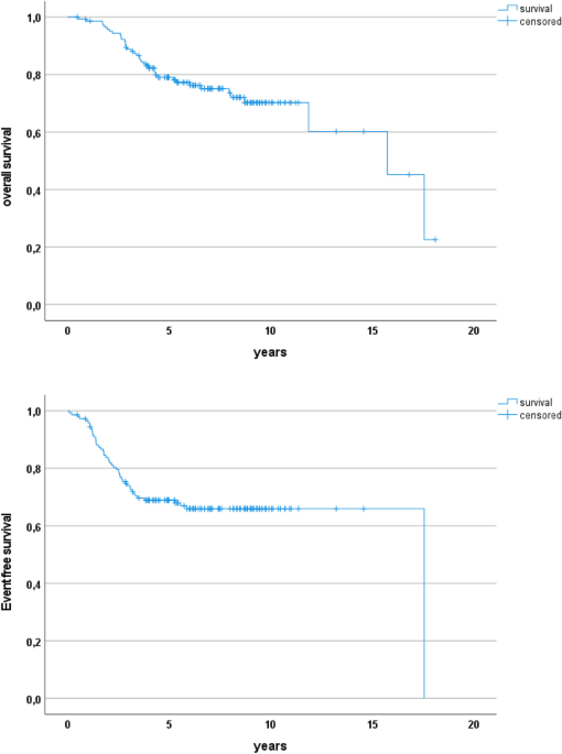
Scheer, Monika; Hallmen, Erika; Vokuhl, Christian; Fuchs, Jörg; Tunn, Per-Ulf; Münter, Marc; Timmermann, Beate; Bauer, Sebastian; Henssen, Anton George; Kazanowska, Bernarda; Niggli, Felix; Ladenstein, Ruth; Ljungman, Gustaf; Eggert, Angelika; Klingebiel, Thomas; and, Ewa Koscielniak
Pre-operative radiotherapy is associated with superior local relapse-free survival in advanced synovial sarcoma Journal Article
In: J Cancer Res Clin Oncol, vol. 149, no. 5, pp. 1717–1731, 2023, ISSN: 1432-1335.
@article{pmid35687182,

title = {Pre-operative radiotherapy is associated with superior local relapse-free survival in advanced synovial sarcoma},

author = {Monika Scheer and Erika Hallmen and Christian Vokuhl and Jörg Fuchs and Per-Ulf Tunn and Marc Münter and Beate Timmermann and Sebastian Bauer and Anton George Henssen and Bernarda Kazanowska and Felix Niggli and Ruth Ladenstein and Gustaf Ljungman and Angelika Eggert and Thomas Klingebiel and Ewa Koscielniak and },

doi = {10.1007/s00432-022-04051-9},

issn = {1432-1335},

year  = {2023},

date = {2023-05-01},

journal = {J Cancer Res Clin Oncol},

volume = {149},

number = {5},

pages = {1717--1731},

abstract = {PURPOSE: Optimization of local therapies in synovial sarcoma (SS) considered unresectable at diagnosis is needed. We evaluated the effects of neoadjuvant versus adjuvant radiation versus surgery only on long-term outcomes.nnMETHODS: Patients with macroscopic SS tumors before chemotherapy (IRS-group-III) in the trials CWS-81, CWS-86, CWS-91, CWS-96, CWS-2002-P and SoTiSaR-registry were analyzed. Local therapies were scheduled after 3 neoadjuvant chemotherapy cycles.nnRESULTS: Median age of 145 patients was 14.5 years. 106 survivors had median follow-up of 7.0 years. Tumor site was 96 extremities, 19 head-neck, 16 shoulder/hip, 14 trunk. Tumors were < 3 cm in 16, 3-5 cm in 28, 5-10 cm in 55, > 10 cm in 34 patients. In a secondary resection during chemotherapy, R0-status was accomplished in 82, R1 in 30, R2 in 21 (12 missing). Radiotherapy was administered to 115 (R0 61, R1 29, R2 20, missing 5), thereof 57 before and 52 after tumor resection. 23 were treated with surgery only. For all patients, 5 year event-free (EFS) and overall survival (OS) was 68.9% ± 7.6 (95%CI) and 79.1% ± 6.9. To establish independent significance, tumor site, size, surgical results and sequencing of local therapies were analyzed in a Cox regression analysis. Variables associated with EFS and OS are site, size and sequencing of local therapies. Variables associated with local recurrence are site, surgical results and sequencing of local therapies. The only variable associated with suffering metastatic recurrence is tumor size.nnCONCLUSION: Differences in sequencing of local therapy procedures are independently associated with outcomes. Best local control is achieved when tumors are irradiated pre-operatively and undergo R0 or R1 resection thereafter.},

keywords = {},

pubstate = {published},

tppubtype = {article}

}

González, Rocío Chamorro; Conrad, Thomas; Stöber, Maja C; Xu, Robin; Giurgiu, Mădălina; Rodriguez-Fos, Elias; Kasack, Katharina; Brückner, Lotte; van Leen, Eric; Helmsauer, Konstantin; Garcia, Heathcliff Dorado; Stefanova, Maria E; Hung, King L; Bei, Yi; Schmelz, Karin; Lodrini, Marco; Mundlos, Stefan; Chang, Howard Y; Deubzer, Hedwig E; Sauer, Sascha; Eggert, Angelika; Schulte, Johannes H; Schwarz, Roland F; Haase, Kerstin; Koche, Richard P; Henssen, Anton G
Parallel sequencing of extrachromosomal circular DNAs and transcriptomes in single cancer cells Journal Article
In: Nat Genet, vol. 55, no. 5, pp. 880–890, 2023, ISSN: 1546-1718.
@article{pmid37142849,

title = {Parallel sequencing of extrachromosomal circular DNAs and transcriptomes in single cancer cells},

author = {Rocío Chamorro González and Thomas Conrad and Maja C Stöber and Robin Xu and Mădălina Giurgiu and Elias Rodriguez-Fos and Katharina Kasack and Lotte Brückner and Eric van Leen and Konstantin Helmsauer and Heathcliff Dorado Garcia and Maria E Stefanova and King L Hung and Yi Bei and Karin Schmelz and Marco Lodrini and Stefan Mundlos and Howard Y Chang and Hedwig E Deubzer and Sascha Sauer and Angelika Eggert and Johannes H Schulte and Roland F Schwarz and Kerstin Haase and Richard P Koche and Anton G Henssen},

doi = {10.1038/s41588-023-01386-y},

issn = {1546-1718},

year  = {2023},

date = {2023-05-01},

journal = {Nat Genet},

volume = {55},

number = {5},

pages = {880--890},

abstract = {Extrachromosomal DNAs (ecDNAs) are common in cancer, but many questions about their origin, structural dynamics and impact on intratumor heterogeneity are still unresolved. Here we describe single-cell extrachromosomal circular DNA and transcriptome sequencing (scEC&T-seq), a method for parallel sequencing of circular DNAs and full-length mRNA from single cells. By applying scEC&T-seq to cancer cells, we describe intercellular differences in ecDNA content while investigating their structural heterogeneity and transcriptional impact. Oncogene-containing ecDNAs were clonally present in cancer cells and drove intercellular oncogene expression differences. In contrast, other small circular DNAs were exclusive to individual cells, indicating differences in their selection and propagation. Intercellular differences in ecDNA structure pointed to circular recombination as a mechanism of ecDNA evolution. These results demonstrate scEC&T-seq as an approach to systematically characterize both small and large circular DNA in cancer cells, which will facilitate the analysis of these DNA elements in cancer and beyond.},

keywords = {},

pubstate = {published},

tppubtype = {article}

}
Rausch, Tobias; Snajder, Rene; Leger, Adrien; Simovic, Milena; Giurgiu, Mădălina; Villacorta, Laura; Henssen, Anton G; Fröhling, Stefan; Stegle, Oliver; Birney, Ewan; Bonder, Marc Jan; Ernst, Aurelie; Korbel, Jan O
Long-read sequencing of diagnosis and post-therapy medulloblastoma reveals complex rearrangement patterns and epigenetic signatures Journal Article
In: Cell Genom, vol. 3, no. 4, pp. 100281, 2023, ISSN: 2666-979X.
@article{pmid37082141,

title = {Long-read sequencing of diagnosis and post-therapy medulloblastoma reveals complex rearrangement patterns and epigenetic signatures},

author = {Tobias Rausch and Rene Snajder and Adrien Leger and Milena Simovic and Mădălina Giurgiu and Laura Villacorta and Anton G Henssen and Stefan Fröhling and Oliver Stegle and Ewan Birney and Marc Jan Bonder and Aurelie Ernst and Jan O Korbel},

doi = {10.1016/j.xgen.2023.100281},

issn = {2666-979X},

year  = {2023},

date = {2023-04-01},

journal = {Cell Genom},

volume = {3},

number = {4},

pages = {100281},

abstract = {Cancer genomes harbor a broad spectrum of structural variants (SVs) driving tumorigenesis, a relevant subset of which escape discovery using short-read sequencing. We employed Oxford Nanopore Technologies (ONT) long-read sequencing in a paired diagnostic and post-therapy medulloblastoma to unravel the haplotype-resolved somatic genetic and epigenetic landscape. We assembled complex rearrangements, including a 1.55-Mbp chromothripsis event, and we uncover a complex SV pattern termed templated insertion (TI) thread, characterized by short (mostly <1 kb) insertions showing prevalent self-concatenation into highly amplified structures of up to 50 kbp in size. TI threads occur in 3% of cancers, with a prevalence up to 74% in liposarcoma, and frequent colocalization with chromothripsis. We also perform long-read-based methylome profiling and discover allele-specific methylation (ASM) effects, complex rearrangements exhibiting differential methylation, and differential promoter methylation in cancer-driver genes. Our study shows the advantage of long-read sequencing in the discovery and characterization of complex somatic rearrangements.},

keywords = {},

pubstate = {published},

tppubtype = {article}

}
Danielli, Sara G; Porpiglia, Ermelinda; Micheli, Andrea J De; Navarro, Natalia; Zellinger, Michael J; Bechtold, Ingrid; Kisele, Samanta; Volken, Larissa; Ngo, Quy A; Marques, Joana G; Kasper, Stephanie; Bode, Peter K; Henssen, Anton G; Gürgen, Dennis; Delattre, Olivier; Surdez, Didier; Roma, Josep; Bühlmann, Peter; Blau, Helen M; Wachtel, Marco; Schäfer, Beat W
Single-cell profiling of alveolar rhabdomyosarcoma reveals RAS pathway inhibitors as cell-fate hijackers with therapeutic relevance Journal Article
In: Sci Adv, vol. 9, no. 6, pp. eade9238, 2023, ISSN: 2375-2548.
@article{pmid36753540,

title = {Single-cell profiling of alveolar rhabdomyosarcoma reveals RAS pathway inhibitors as cell-fate hijackers with therapeutic relevance},

author = {Sara G Danielli and Ermelinda Porpiglia and Andrea J De Micheli and Natalia Navarro and Michael J Zellinger and Ingrid Bechtold and Samanta Kisele and Larissa Volken and Quy A Ngo and Joana G Marques and Stephanie Kasper and Peter K Bode and Anton G Henssen and Dennis Gürgen and Olivier Delattre and Didier Surdez and Josep Roma and Peter Bühlmann and Helen M Blau and Marco Wachtel and Beat W Schäfer},

doi = {10.1126/sciadv.ade9238},

issn = {2375-2548},

year  = {2023},

date = {2023-02-01},

journal = {Sci Adv},

volume = {9},

number = {6},

pages = {eade9238},

abstract = {Rhabdomyosarcoma (RMS) is a group of pediatric cancers with features of developing skeletal muscle. The cellular hierarchy and mechanisms leading to developmental arrest remain elusive. Here, we combined single-cell RNA sequencing, mass cytometry, and high-content imaging to resolve intratumoral heterogeneity of patient-derived primary RMS cultures. We show that the aggressive alveolar RMS (aRMS) subtype contains plastic muscle stem-like cells and cycling progenitors that drive tumor growth, and a subpopulation of differentiated cells that lost its proliferative potential and correlates with better outcomes. While chemotherapy eliminates cycling progenitors, it enriches aRMS for muscle stem-like cells. We screened for drugs hijacking aRMS toward clinically favorable subpopulations and identified a combination of RAF and MEK inhibitors that potently induces myogenic differentiation and inhibits tumor growth. Overall, our work provides insights into the developmental states underlying aRMS aggressiveness, chemoresistance, and progression and identifies the RAS pathway as a promising therapeutic target.},

keywords = {},

pubstate = {published},

tppubtype = {article}

}
Launspach, Michael; Temel, Dennis; Ohlendorf, Emily; Zirngibl, Felix; Materne, Bianca; Oevermann, Lena; Deubzer, Hedwig E; Henssen, Anton G; Künkele, Annette; Hundsdörfer, Patrick; von Bernuth, Horst; Pruß, Axel; Eggert, Angelika; von Stackelberg, Arend; Lang, Peter; Schulte, Johannes H
2023, ISSN: 1592-8721.
@misc{pmid36134454,

title = {Rituximab therapy after pediatric hematopoietic stem cell transplantation can cause prolonged B-cell impairment and increases the risk for infections - a retrospective matched cohort study},

author = {Michael Launspach and Dennis Temel and Emily Ohlendorf and Felix Zirngibl and Bianca Materne and Lena Oevermann and Hedwig E Deubzer and Anton G Henssen and Annette Künkele and Patrick Hundsdörfer and Horst von Bernuth and Axel Pruß and Angelika Eggert and Arend von Stackelberg and Peter Lang and Johannes H Schulte},

doi = {10.3324/haematol.2022.281134},

issn = {1592-8721},

year  = {2023},

date = {2023-01-01},

journal = {Haematologica},

volume = {108},

number = {1},

pages = {267--272},

keywords = {},

pubstate = {published},

tppubtype = {misc}

}
Pecorino, Lauren T; Verhaak, Roel G W; Henssen, Anton; Mischel, Paul S
Extrachromosomal DNA (ecDNA): an origin of tumor heterogeneity, genomic remodeling, and drug resistance Journal Article
In: Biochem Soc Trans, vol. 50, no. 6, pp. 1911–1920, 2022, ISSN: 1470-8752.
@article{pmid36355400,

title = {Extrachromosomal DNA (ecDNA): an origin of tumor heterogeneity, genomic remodeling, and drug resistance},

author = {Lauren T Pecorino and Roel G W Verhaak and Anton Henssen and Paul S Mischel},

doi = {10.1042/BST20221045},

issn = {1470-8752},

year  = {2022},

date = {2022-12-01},

journal = {Biochem Soc Trans},

volume = {50},

number = {6},

pages = {1911--1920},

abstract = {The genome of cancer cells contains circular extrachromosomal DNA (ecDNA) elements not found in normal cells. Analysis of clinical samples reveal they are common in most cancers and their presence indicates poor prognosis. They often contain enhancers and driver oncogenes that are highly expressed. The circular ecDNA topology leads to an open chromatin conformation and generates new gene regulatory interactions, including with distal enhancers. The absence of centromeres leads to random distribution of ecDNAs during cell division and genes encoded on them are transmitted in a non-mendelian manner. ecDNA can integrate into and exit from chromosomal DNA. The numbers of specific ecDNAs can change in response to treatment. This dynamic ability to remodel the cancer genome challenges long-standing fundamentals, providing new insights into tumor heterogeneity, cancer genome remodeling, and drug resistance.},

keywords = {},

pubstate = {published},

tppubtype = {article}

}
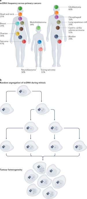
Yi, Eunhee; González, Rocío Chamorro; Henssen, Anton G; Verhaak, Roel G W
Extrachromosomal DNA amplifications in cancer Journal Article
In: Nat Rev Genet, vol. 23, no. 12, pp. 760–771, 2022, ISSN: 1471-0064.
@article{pmid35953594,

title = {Extrachromosomal DNA amplifications in cancer},

author = {Eunhee Yi and Rocío Chamorro González and Anton G Henssen and Roel G W Verhaak},

doi = {10.1038/s41576-022-00521-5},

issn = {1471-0064},

year  = {2022},

date = {2022-12-01},

journal = {Nat Rev Genet},

volume = {23},

number = {12},

pages = {760--771},

abstract = {Extrachromosomal DNA (ecDNA) amplification is an important driver alteration in cancer. It has been observed in most cancer types and is associated with worse patient outcome. The functional impact of ecDNA has been linked to its unique properties, such as its circular structure that is associated with altered chromatinization and epigenetic regulatory landscape, as well as its ability to randomly segregate during cell division, which fuels intercellular copy number heterogeneity. Recent investigations suggest that ecDNA is structurally more complex than previously anticipated and that it localizes to specialized nuclear bodies (hubs) and can act in trans as an enhancer for genes on other ecDNAs or chromosomes. In this Review, we synthesize what is currently known about how ecDNA is generated and how its genetic and epigenetic architecture affects proto-oncogene deregulation in cancer. We discuss how recently identified ecDNA functions may impact oncogenesis but also serve as new therapeutic vulnerabilities in cancer.},

keywords = {},

pubstate = {published},

tppubtype = {article}

}

Hung, King L; Luebeck, Jens; Dehkordi, Siavash R; Colón, Caterina I; Li, Rui; Wong, Ivy Tsz-Lo; Coruh, Ceyda; Dharanipragada, Prashanthi; Lomeli, Shirley H; Weiser, Natasha E; Moriceau, Gatien; Zhang, Xiao; Bailey, Chris; Houlahan, Kathleen E; Yang, Wenting; González, Rocío Chamorro; Swanton, Charles; Curtis, Christina; Jamal-Hanjani, Mariam; Henssen, Anton G; Law, Julie A; Greenleaf, William J; Lo, Roger S; Mischel, Paul S; Bafna, Vineet; Chang, Howard Y
Targeted profiling of human extrachromosomal DNA by CRISPR-CATCH Journal Article
In: Nat Genet, vol. 54, no. 11, pp. 1746–1754, 2022, ISSN: 1546-1718.
@article{pmid36253572,

title = {Targeted profiling of human extrachromosomal DNA by CRISPR-CATCH},

author = {King L Hung and Jens Luebeck and Siavash R Dehkordi and Caterina I Colón and Rui Li and Ivy Tsz-Lo Wong and Ceyda Coruh and Prashanthi Dharanipragada and Shirley H Lomeli and Natasha E Weiser and Gatien Moriceau and Xiao Zhang and Chris Bailey and Kathleen E Houlahan and Wenting Yang and Rocío Chamorro González and Charles Swanton and Christina Curtis and Mariam Jamal-Hanjani and Anton G Henssen and Julie A Law and William J Greenleaf and Roger S Lo and Paul S Mischel and Vineet Bafna and Howard Y Chang},

doi = {10.1038/s41588-022-01190-0},

issn = {1546-1718},

year  = {2022},

date = {2022-11-01},

journal = {Nat Genet},

volume = {54},

number = {11},

pages = {1746--1754},

abstract = {Extrachromosomal DNA (ecDNA) is a common mode of oncogene amplification but is challenging to analyze. Here, we adapt CRISPR-CATCH, in vitro CRISPR-Cas9 treatment and pulsed field gel electrophoresis of agarose-entrapped genomic DNA, previously developed for bacterial chromosome segments, to isolate megabase-sized human ecDNAs. We demonstrate strong enrichment of ecDNA molecules containing EGFR, FGFR2 and MYC from human cancer cells and NRAS ecDNA from human metastatic melanoma with acquired therapeutic resistance. Targeted enrichment of ecDNA versus chromosomal DNA enabled phasing of genetic variants, identified the presence of an EGFRvIII mutation exclusively on ecDNAs and supported an excision model of ecDNA genesis in a glioblastoma model. CRISPR-CATCH followed by nanopore sequencing enabled single-molecule ecDNA methylation profiling and revealed hypomethylation of the EGFR promoter on ecDNAs. We distinguished heterogeneous ecDNA species within the same sample by size and sequence with base-pair resolution and discovered functionally specialized ecDNAs that amplify select enhancers or oncogene-coding sequences.},

keywords = {},

pubstate = {published},

tppubtype = {article}

}
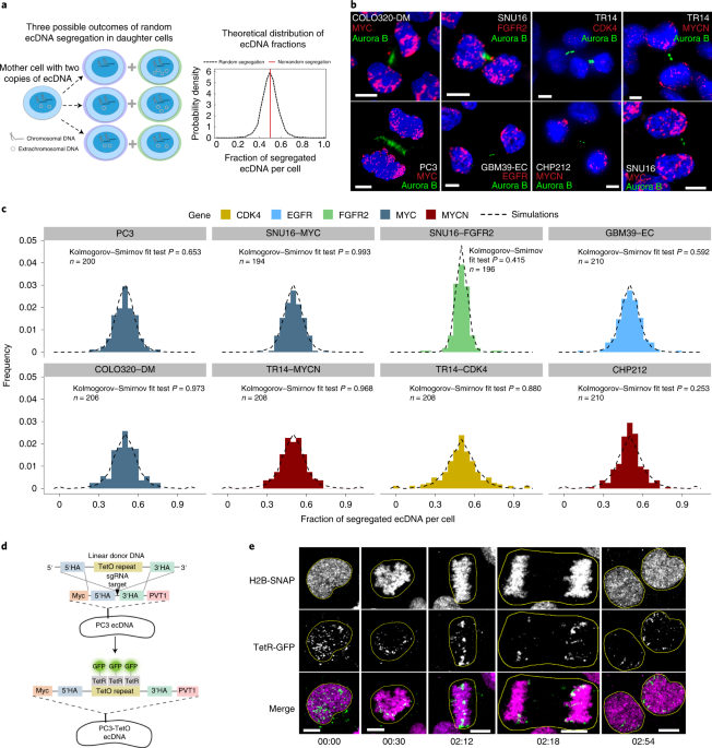
Lange, Joshua T; Rose, John C; Chen, Celine Y; Pichugin, Yuriy; Xie, Liangqi; Tang, Jun; Hung, King L; Yost, Kathryn E; Shi, Quanming; Erb, Marcella L; Rajkumar, Utkrisht; Wu, Sihan; Taschner-Mandl, Sabine; Bernkopf, Marie; Swanton, Charles; Liu, Zhe; Huang, Weini; Chang, Howard Y; Bafna, Vineet; Henssen, Anton G; Werner, Benjamin; Mischel, Paul S
The evolutionary dynamics of extrachromosomal DNA in human cancers Journal Article
In: Nat Genet, vol. 54, no. 10, pp. 1527–1533, 2022, ISSN: 1546-1718.
@article{pmid36123406,

title = {The evolutionary dynamics of extrachromosomal DNA in human cancers},

author = {Joshua T Lange and John C Rose and Celine Y Chen and Yuriy Pichugin and Liangqi Xie and Jun Tang and King L Hung and Kathryn E Yost and Quanming Shi and Marcella L Erb and Utkrisht Rajkumar and Sihan Wu and Sabine Taschner-Mandl and Marie Bernkopf and Charles Swanton and Zhe Liu and Weini Huang and Howard Y Chang and Vineet Bafna and Anton G Henssen and Benjamin Werner and Paul S Mischel},

doi = {10.1038/s41588-022-01177-x},

issn = {1546-1718},

year  = {2022},

date = {2022-10-01},

journal = {Nat Genet},

volume = {54},

number = {10},

pages = {1527--1533},

abstract = {Oncogene amplification on extrachromosomal DNA (ecDNA) is a common event, driving aggressive tumor growth, drug resistance and shorter survival. Currently, the impact of nonchromosomal oncogene inheritance-random identity by descent-is poorly understood. Also unclear is the impact of ecDNA on somatic variation and selection. Here integrating theoretical models of random segregation, unbiased image analysis, CRISPR-based ecDNA tagging with live-cell imaging and CRISPR-C, we demonstrate that random ecDNA inheritance results in extensive intratumoral ecDNA copy number heterogeneity and rapid adaptation to metabolic stress and targeted treatment. Observed ecDNAs benefit host cell survival or growth and can change within a single cell cycle. ecDNA inheritance can predict, a priori, some of the aggressive features of ecDNA-containing cancers. These properties are facilitated by the ability of ecDNA to rapidly adapt genomes in a way that is not possible through chromosomal oncogene amplification. These results show how the nonchromosomal random inheritance pattern of ecDNA contributes to poor outcomes for patients with cancer.},

keywords = {},

pubstate = {published},

tppubtype = {article}

}
Schubert, Nil A; Chen, Celine Y; Rodríguez, Ana; Koster, Jan; Dowless, Michele; Pfister, Stefan M; Shields, David J; Stancato, Louis F; Vassal, Gilles; Caron, Hubert N; van den Boogaard, Marlinde L; Henssen, Anton G; Molenaar, Jan J
Target actionability review to evaluate CDK4/6 as a therapeutic target in paediatric solid and brain tumours Journal Article
In: Eur J Cancer, vol. 170, pp. 196–208, 2022, ISSN: 1879-0852.
@article{pmid35671543,

title = {Target actionability review to evaluate CDK4/6 as a therapeutic target in paediatric solid and brain tumours},

author = {Nil A Schubert and Celine Y Chen and Ana Rodríguez and Jan Koster and Michele Dowless and Stefan M Pfister and David J Shields and Louis F Stancato and Gilles Vassal and Hubert N Caron and Marlinde L van den Boogaard and Anton G Henssen and Jan J Molenaar},

doi = {10.1016/j.ejca.2022.04.028},

issn = {1879-0852},

year  = {2022},

date = {2022-07-01},

journal = {Eur J Cancer},

volume = {170},

pages = {196--208},

abstract = {BACKGROUND: Childhood cancer is still a leading cause of death around the world. To improve outcomes, there is an urgent need for tailored treatment. The systematic evaluation of existing preclinical data can provide an overview of what is known and identify gaps in the current knowledge. Here, we applied the target actionability review (TAR) methodology to assess the strength and weaknesses of available scientific literature on CDK4/6 as a therapeutic target in paediatric solid and brain tumours by structured critical appraisal.nnMETHODS: Using relevant search terms in PubMed, a list of original publications investigating CDK4/6 in paediatric solid tumour types was identified based on relevancy criteria. Each publication was annotated for the tumour type and categorised into separate proof-of-concept (PoC) data modules. Based on rubrics, quality and experimental outcomes were scored independently by two reviewers. A third reviewer evaluated and adjudicated score discrepancies. Scores for each PoC module were averaged for each tumour type and visualised in a heatmap matrix in the publicly available R2 data portal.nnRESULTS AND CONCLUSIONS: This CDK4/6 TAR, generated by analysis of 151 data entries from 71 publications, showed frequent genomic aberrations of CDK4/6 in rhabdomyosarcoma, osteosarcoma, high-grade glioma, medulloblastoma, and neuroblastoma. However, a clear correlation between CDK4/6 aberrations and compound efficacy is not coming forth from the literature. Our analysis indicates that several paediatric indications would need (further) preclinical evaluation to allow for better recommendations, especially regarding the dependence of tumours on CDK4/6, predictive biomarkers, resistance mechanisms, and combination strategies. Nevertheless, our TAR heatmap provides support for the relevance of CDK4/6 inhibition in Ewing sarcoma, medulloblastoma, malignant peripheral nerve sheath tumour and to a lesser extent neuroblastoma, rhabdomyosarcoma, rhabdoid tumour and high-grade glioma. The interactive heatmap is accessible through R2 [r2platform.com/TAR/CDK4_6].},

keywords = {},

pubstate = {published},

tppubtype = {article}

}
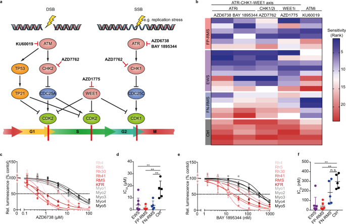
García, Heathcliff Dorado; Pusch, Fabian; Bei, Yi; von Stebut, Jennifer; Ibáñez, Glorymar; Guillan, Kristina; Imami, Koshi; Gürgen, Dennis; Rolff, Jana; Helmsauer, Konstantin; Meyer-Liesener, Stephanie; Timme, Natalie; Bardinet, Victor; González, Rocío Chamorro; MacArthur, Ian C; Chen, Celine Y; Schulz, Joachim; Wengner, Antje M; Furth, Christian; Lala, Birgit; Eggert, Angelika; Seifert, Georg; Hundsoerfer, Patrick; Kirchner, Marieluise; Mertins, Philipp; Selbach, Matthias; Lissat, Andrej; Dubois, Frank; Horst, David; Schulte, Johannes H; Spuler, Simone; You, Daoqi; Cruz, Filemon Dela; Kung, Andrew L; Haase, Kerstin; DiVirgilio, Michela; Scheer, Monika; Ortiz, Michael V; Henssen, Anton G
Therapeutic targeting of ATR in alveolar rhabdomyosarcoma Journal Article
In: Nat Commun, vol. 13, no. 1, pp. 4297, 2022, ISSN: 2041-1723.
@article{pmid35879366,

title = {Therapeutic targeting of ATR in alveolar rhabdomyosarcoma},

author = {Heathcliff Dorado García and Fabian Pusch and Yi Bei and Jennifer von Stebut and Glorymar Ibáñez and Kristina Guillan and Koshi Imami and Dennis Gürgen and Jana Rolff and Konstantin Helmsauer and Stephanie Meyer-Liesener and Natalie Timme and Victor Bardinet and Rocío Chamorro González and Ian C MacArthur and Celine Y Chen and Joachim Schulz and Antje M Wengner and Christian Furth and Birgit Lala and Angelika Eggert and Georg Seifert and Patrick Hundsoerfer and Marieluise Kirchner and Philipp Mertins and Matthias Selbach and Andrej Lissat and Frank Dubois and David Horst and Johannes H Schulte and Simone Spuler and Daoqi You and Filemon Dela Cruz and Andrew L Kung and Kerstin Haase and Michela DiVirgilio and Monika Scheer and Michael V Ortiz and Anton G Henssen},

doi = {10.1038/s41467-022-32023-7},

issn = {2041-1723},

year  = {2022},

date = {2022-07-01},

journal = {Nat Commun},

volume = {13},

number = {1},

pages = {4297},

abstract = {Despite advances in multi-modal treatment approaches, clinical outcomes of patients suffering from PAX3-FOXO1 fusion oncogene-expressing alveolar rhabdomyosarcoma (ARMS) remain dismal. Here we show that PAX3-FOXO1-expressing ARMS cells are sensitive to pharmacological ataxia telangiectasia and Rad3 related protein (ATR) inhibition. Expression of PAX3-FOXO1 in muscle progenitor cells is not only sufficient to increase sensitivity to ATR inhibition, but PAX3-FOXO1-expressing rhabdomyosarcoma cells also exhibit increased sensitivity to structurally diverse inhibitors of ATR. Mechanistically, ATR inhibition leads to replication stress exacerbation, decreased BRCA1 phosphorylation and reduced homologous recombination-mediated DNA repair pathway activity. Consequently, ATR inhibitor treatment increases sensitivity of ARMS cells to PARP1 inhibition in vitro, and combined treatment with ATR and PARP1 inhibitors induces complete regression of primary patient-derived ARMS xenografts in vivo. Lastly, a genome-wide CRISPR activation screen (CRISPRa) in combination with transcriptional analyses of ATR inhibitor resistant ARMS cells identifies the RAS-MAPK pathway and its targets, the FOS gene family, as inducers of resistance to ATR inhibition. Our findings provide a rationale for upcoming biomarker-driven clinical trials of ATR inhibitors in patients suffering from ARMS.},

keywords = {},

pubstate = {published},

tppubtype = {article}

}
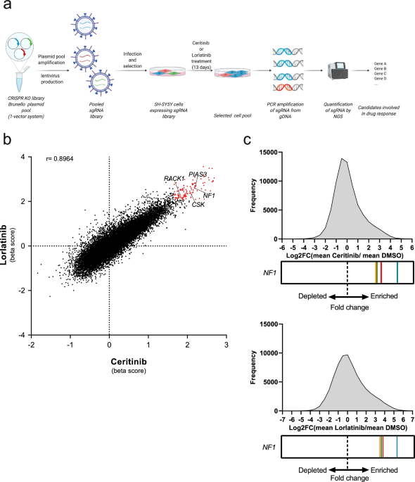
Berlak, Mareike; Tucker, Elizabeth; Dorel, Mathurin; Winkler, Annika; McGearey, Aleixandria; Rodriguez-Fos, Elias; da Costa, Barbara Martins; Barker, Karen; Fyle, Elicia; Calton, Elizabeth; Eising, Selma; Ober, Kim; Hughes, Deborah; Koutroumanidou, Eleni; Carter, Paul; Stankunaite, Reda; Proszek, Paula; Jain, Neha; Rosswog, Carolina; Dorado-Garcia, Heathcliff; Molenaar, Jan Jasper; Hubank, Mike; Barone, Giuseppe; Anderson, John; Lang, Peter; Deubzer, Hedwig Elisabeth; Künkele, Annette; Fischer, Matthias; Eggert, Angelika; Kloft, Charlotte; Henssen, Anton George; Boettcher, Michael; Hertwig, Falk; Blüthgen, Nils; Chesler, Louis; Schulte, Johannes Hubertus
Mutations in ALK signaling pathways conferring resistance to ALK inhibitor treatment lead to collateral vulnerabilities in neuroblastoma cells Journal Article
In: Mol Cancer, vol. 21, no. 1, pp. 126, 2022, ISSN: 1476-4598.
@article{pmid35689207,

title = {Mutations in ALK signaling pathways conferring resistance to ALK inhibitor treatment lead to collateral vulnerabilities in neuroblastoma cells},

author = {Mareike Berlak and Elizabeth Tucker and Mathurin Dorel and Annika Winkler and Aleixandria McGearey and Elias Rodriguez-Fos and Barbara Martins da Costa and Karen Barker and Elicia Fyle and Elizabeth Calton and Selma Eising and Kim Ober and Deborah Hughes and Eleni Koutroumanidou and Paul Carter and Reda Stankunaite and Paula Proszek and Neha Jain and Carolina Rosswog and Heathcliff Dorado-Garcia and Jan Jasper Molenaar and Mike Hubank and Giuseppe Barone and John Anderson and Peter Lang and Hedwig Elisabeth Deubzer and Annette Künkele and Matthias Fischer and Angelika Eggert and Charlotte Kloft and Anton George Henssen and Michael Boettcher and Falk Hertwig and Nils Blüthgen and Louis Chesler and Johannes Hubertus Schulte},

doi = {10.1186/s12943-022-01583-z},

issn = {1476-4598},

year  = {2022},

date = {2022-06-01},

journal = {Mol Cancer},

volume = {21},

number = {1},

pages = {126},

abstract = {BACKGROUND: Development of resistance to targeted therapies has tempered initial optimism that precision oncology would improve poor outcomes for cancer patients. Resistance mechanisms, however, can also confer new resistance-specific vulnerabilities, termed collateral sensitivities. Here we investigated anaplastic lymphoma kinase (ALK) inhibitor resistance in neuroblastoma, a childhood cancer frequently affected by activating ALK alterations.nnMETHODS: Genome-wide forward genetic CRISPR-Cas9 based screens were performed to identify genes associated with ALK inhibitor resistance in neuroblastoma cell lines. Furthermore, the neuroblastoma cell line NBLW-R was rendered resistant by continuous exposure to ALK inhibitors. Genes identified to be associated with ALK inhibitor resistance were further investigated by generating suitable cell line models. In addition, tumor and liquid biopsy samples of four patients with ALK-mutated neuroblastomas before ALK inhibitor treatment and during tumor progression under treatment were genomically profiled.nnRESULTS: Both genome-wide CRISPR-Cas9-based screens and preclinical spontaneous ALKi resistance models identified NF1 loss and activating NRAS mutations to confer resistance to chemically diverse ALKi. Moreover, human neuroblastomas recurrently developed de novo loss of NF1 and activating RAS mutations after ALKi treatment, leading to therapy resistance. Pathway-specific perturbations confirmed that NF1 loss and activating RAS mutations lead to RAS-MAPK signaling even in the presence of ALKi. Intriguingly, NF1 loss rendered neuroblastoma cells hypersensitive to MEK inhibition.nnCONCLUSIONS: Our results provide a clinically relevant mechanistic model of ALKi resistance in neuroblastoma and highlight new clinically actionable collateral sensitivities in resistant cells.},

keywords = {},

pubstate = {published},

tppubtype = {article}

}
Thorwarth, Anne; Haase, Kerstin; Röefzaad, Claudia; Pajtler, Kristian W; Schramm, Kathrin; Hauptmann, Kathrin; Behnke, Anke; Vokuhl, Christian; Elgeti, Thomas; Gratopp, Alexander; Schulte, Johannes H; Scheer, Monika; Driever, Pablo Hernáiz; Nysom, Karsten; Eggert, Angelika; Henssen, Anton G; Deubzer, Hedwig E
Genomic Evolution and Personalized Therapy of an Infantile Fibrosarcoma Harboring an Oncogenic Fusion Journal Article
In: JCO Precis Oncol, vol. 6, pp. e2100283, 2022, ISSN: 2473-4284.
@article{pmid35613412,

title = {Genomic Evolution and Personalized Therapy of an Infantile Fibrosarcoma Harboring an  Oncogenic Fusion},

author = {Anne Thorwarth and Kerstin Haase and Claudia Röefzaad and Kristian W Pajtler and Kathrin Schramm and Kathrin Hauptmann and Anke Behnke and Christian Vokuhl and Thomas Elgeti and Alexander Gratopp and Johannes H Schulte and Monika Scheer and Pablo Hernáiz Driever and Karsten Nysom and Angelika Eggert and Anton G Henssen and Hedwig E Deubzer},

doi = {10.1200/PO.21.00283},

issn = {2473-4284},

year  = {2022},

date = {2022-05-01},

journal = {JCO Precis Oncol},

volume = {6},

pages = {e2100283},

keywords = {},

pubstate = {published},

tppubtype = {article}

}
Henssen, Anton G
Shedding Light on ecDNA Behavior Using CRISPR-Based Live-Cell Imaging Journal Article
In: Cancer Discov, vol. 12, no. 2, pp. 293–295, 2022, ISSN: 2159-8290.
@article{pmid35140176,

title = {Shedding Light on ecDNA Behavior Using CRISPR-Based Live-Cell Imaging},

author = {Anton G Henssen},

doi = {10.1158/2159-8290.CD-21-1525},

issn = {2159-8290},

year  = {2022},

date = {2022-02-01},

journal = {Cancer Discov},

volume = {12},

number = {2},

pages = {293--295},

abstract = {Extrachromosomal oncogene amplification on extrachromosomal DNA (ecDNA) has emerged as a hallmark of many cancers. In this issue, Yi and colleagues developed a CRISPR-based method for imaging ecDNA in live cells, termed ecTag. Using ecTag, the authors reveal important features of ecDNA in cancer cells such as their random mitotic segregation and clustering into transcriptionally active hubs after mitosis..},

keywords = {},

pubstate = {published},

tppubtype = {article}

}